Download
1 / 40
400 likes | 423 Views
This article explores the process of antigen capture and presentation by antigen-presenting cells (APCs) in the context of major histocompatibility complex (MHC) molecules. It discusses the role of different APCs, such as dendritic cells, macrophages, and B lymphocytes, in activating T cells and the importance of MHC molecules in presenting antigen peptides. The article also highlights the general properties of APCs and the polymorphic nature of MHC genes.

E N D
Antigen Presentation and Major Histocompatibility Complex Molecules
ANTIGEN CAPTURE AND THE FUNCTIONS OF ANTIGEN-PRESENTING CELLS
Antigens are captured from their site of entry and concentrated in peripheral (secondary) lymphoid organs through which naive T cells circulate constantly. • Lymphocytes recognize and respond to cell-associated antigens and not to soluble, cell-free antigens. • The antigen receptors of most T cells recognize only peptides displayed by MHC molecules on the surface of antigen-presenting cells (APCs).
Dendritic cells are the most efficient APCs for initiating primary responses by activating naive T cells, and macrophages and B lymphocytes present antigens to helper T cells in the effector phase of cell-mediated immunity and in humoral Irs, respectively. • All nucleated cells can present class I–associated peptides, derived from cytosolic proteins such as viral and tumor antigens, to CD8+ T cells.
CD4+ helper T lymphocytes recognize antigens in association with class II MHC molecules, and CD8+ CTLs recognize antigens in association with class I MHC molecules. • APCs capture protein antigens, process them, and display MHC–associated peptides to T cells. • A single T cell can recognize a specific peptide displayed by only one of the large number of different MHC molecules that exist. This phenomenon is called MHC restriction.
MHC-associated peptides contain some residues that anchor them into pockets in the cleft of the MHC molecule and other residues that are recognized by T cell antigen receptors. (Anchor residue determine MHC specificity to the antigen) • MHC residues that may vary among individuals (polymorphic residues) are also recognized by the T cell receptor. Thus, T cells see both peptide antigens and MHC molecules.
General Properties of Antigen-Presenting Cells • Different cell types function as antigen-presenting cells to activate naive T cells or previously differentiated effector T cells. • DCs are the most effective APCs for activating naive T cells and therefore for initiating T cell responses. Macrophages and B lymphocytes also function as APCs, but mostly for previously activated CD4+ helper T cells rather than for naive T cells.
Several properties of DCs make them the most efficient APCs for initiating primary T cell responses • DCs are strategically located at the common sites of entry of microbes and foreign antigens (in epithelia) and in tissues that may be colonized by microbes. • DCs express receptors that enable them to capture and respond to microbes. • DCs migrate from epithelia and tissues via lymphatics, preferentially into the T cell zones of lymph nodes, and naive T lymphocytes also circulate through the same regions of the lymph nodes. • Mature DCs express high levels of peptide-MHC complexes, costimulators, and cytokines, all of which are needed to activate naive T lymphocytes.
The MHC locus contains two types of polymorphic MHC genes, the class I and class II MHC genes, which encode two groups of structurally distinct but homologous proteins, and other nonpolymorphic genes whose products are involved in antigen presentation • Class I and class II MHC genes are the most polymorphic genes present in any mammalian genome. These genes are located at chromosome 6.
MHC genes are codominantly expressed in each individual. • The set of MHC alleles present on each chromosome is called an MHC haplotype. For instance, an HLA haplotype of an individual could be HLA-A2, B5, DR3, and so on (using the simpler nomenclature for HLA alleles). • Class I molecules are expressed on virtually all nucleated cells, whereas class II molecules are expressed only on dendritic cells, B lymphocytes, macrophages, thymic epithelial cells, and a few other cell types.
The expression of MHC molecules is increased by cytokines produced during both innate and adaptive immune responses.
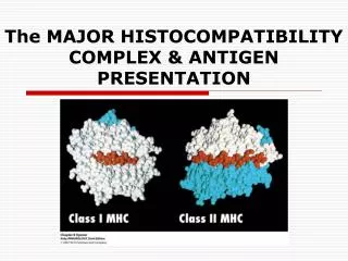
Class I MHC molecules are composed of an α (or heavy) chain in a noncovalent complex with a nonpolymorphic polypeptide called β2-microglobulin. • Class II MHC molecules contain two MHC-encoded polymorphic chains, an α chain and a β chain. • Both classes of MHC molecules consist of an extracellular peptide-binding cleft, a nonpolymorphic Ig-like region, a transmembrane region, and a cytoplasmic region.
The peptide-binding cleft of MHC molecules has α-helical sides and an eight-stranded antiparallel β-pleated sheet floor. • Y The Ig-like domains of class I and class II MHC molecules contain the binding sites for the T cell coreceptors CD8 and CD4, respectively. • The polymorphic residues of MHC molecules are localized to the peptide-binding domain.
The fully assembled class I or II molecule is a trimeric complex consisting of an α chain, β2-microglobulin, and a bound peptide, and stable expression of class I or II molecules on cell surfaces requires the presence of all three components of the complex.
MHC molecules bind only one peptide at a time, and all of the peptides that bind to a particular MHC molecule share common structural motifs. • Every MHC molecule has a broad specificity for peptides and can bind multiple peptides that have common structural features, such as anchor residues.
Characteristics of Peptide-MHC Molecule Interactions • MHC molecules show a broad specificity for peptide binding, in contrast to the fine specificity of antigen recognition by the antigen receptors of lymphocytes. • Each class I or class II MHC molecule has a single peptide-binding cleft that binds one peptide at a time, but each MHC molecule can bind many different peptides.
Characteristics of Peptide-MHC Molecule Interactions • The peptides that bind to MHC molecules share structural features that promote this interaction. • MHC molecules acquire their peptide cargo during their biosynthesis and assembly inside cells. • The association of peptides and MHC molecules is a saturable interaction with a very slow off-rate
Characteristics of Peptide-MHC Molecule Interactions • Very small numbers of peptide-MHC complexes are capable of activating specific T lymphocytes. • The MHC molecules of an individual can bind and display foreign peptides (e.g., those derived from microbial proteins) as well as peptides derived from the proteins of that individual (self antigens).
The Phenomenon of MHC Restriction • This theory arise from examining the recognition of virus-infected cells by virus-specific CTLs in inbred mice. • If a mouse is infected with a virus, CD8+ T cells specific for the virus are activated and differentiate into CTLs in the animal. • When the function of these CTLs is analyzed in vitro, they recognize and kill virus-infected cells only if the infected cells express MHC molecules that are expressed in the animal from which the CTLs were removed.
The Phenomenon of MHC Restriction • Thus, T cells must be specific not only for the antigen but also for MHC molecules, and T cell antigen recognition is restricted by the MHC molecules a T cell sees. • Subsequent studies established that the recognition of antigens by CD8+ CTLs is restricted by class I MHC molecules, and the responses of CD4+ helper T lymphocytes to antigens are restricted by class II MHC molecules.
Antigen processing and presentation • Antigen processing is the conversion of native proteins into MHC-associated peptides. • This process consists of the introduction of exogenous protein antigens into vesicles of APCs or the synthesis of antigens in the cytosol, the proteolytic degradation of these proteins into peptides, the binding of peptides to MHC molecules, and the display of the peptide-MHC complexes on the APC surface for recognition by T cells. • Thus, both extracellular and intracellular proteins are sampled by these antigen processing pathways, and peptides derived from both normal self proteins and foreign proteins are displayed by MHC molecules for surveillance by T lymphocytes.
The Class I MHC Pathway for Processing and Presentation of Cytosolic Proteins • For the class I MHC pathway, protein antigens are degraded in the proteasome, generating peptides that bind to class I MHC molecules. • Most of these antigens are synthesized in the cytosol or introduced into the cytosol from microbes or vesicles. • These peptides are delivered from the cytosol to the ER by an ATP-dependent transporter called TAP.
The Class I MHC Pathway for Processing and Presentation of Cytosolic Proteins • Newly synthesized class I MHC–β2-microglobulin dimers in the ER are associated with the TAP complex and receive peptides transported into the ER. • Stable complexes of class I MHC molecules with bound peptides move out of the ER, through the Golgi complex, to the cell surface.
The Class II MHC Pathway for Presentation of Proteins Degraded in Lysosomes • For the class II MHC pathway, protein antigens are internalized into endosomes, and these proteins are proteolytically cleaved by enzymes in lysosomes and late endosomes. • Newly synthesized class II MHC molecules associated with the invariant chain (Ii) are transported from the ER to the endosomal vesicles. Here the Ii is proteolytically cleaved, and a small peptide remnant of the Ii, called CLIP, is removed from the peptide-binding cleft of the MHC molecule by the DM molecules.
The Class II MHC Pathway for Presentation of Proteins Degraded in Lysosomes • The peptides that were generated from extracellular proteins then bind to the available cleft of the class II MHC molecule, and the trimeric complex (class II MHC α and β chains and peptide) moves to and is displayed on the surface of the cell.
These pathways of MHC-restricted antigen presentation ensure that most of the body’s cells are screened for the possible presence of foreign antigens. • The pathways also ensure that proteins from extracellular microbes preferentially generate peptides bound to class II MHC molecules for recognition by CD4+ helper T cells, which activate effector mechanisms that eliminate extracellular antigens. • Conversely, proteins synthesized by intracellular (cytosolic) microbes generate peptides bound to class I MHC molecules for recognition by CD8+ CTLs, which function to eliminate cells harboring intracellular infections. • The immunogenicity of foreign protein antigens depends on the ability of antigen-processing pathways to generate peptides from the proteins that bind to self MHC molecules.