
Proton NMR Spectroscopy: Basics and Fundamentals
E N D
Presentation Transcript
(NMR) Chapter 3
ʏ ʏ relativistic correction
Because all 12hydrogen atoms in a tetramethylsilane molecule are equivalent, its 1H NMR spectrum consists of a singlet, The chemical shift of this singlet is assigned as δ 0, and all other chemical shifts are determined relative to it.
Initially, there is no difference between spin states in the absence of an external magnetic field present. The presence of the external field will cause a divergence between higher and lower energetic spin states. Energy As the intensity of external field increase so does the separation in the energy widen between spin states. (3) Notice how the negative spin state, , is higher than the positive spin state, .
= chemical shift (Hz) – shift of tetramethylsilane (TMS; 0 Hz) = ppm spectrometer frequency (MHz)
Information from 1H-nmr spectra: • Number of signals: How many different types of hydrogens in the molecule. • Position of signals (chemical shift): What types of hydrogens. • Relative areas under signals (integration): How many hydrogens of each type. • Splitting pattern: How many neighboring hydrogens. Magnetically equivalent hydrogens resonate at the same applied field.
In the presence of an external magnetic field B, the magnetic moments align and they precess around the external field direction with a frequency w0which is called the Larmor frequency. The Larmor frequency is proportional to the gyromagnetic ratio g of the atoms. g is unique for a nucleus
Excitation by RF Energy and Subsequent Relaxation T1 = spin-lattice relaxation time; establishes the z axis equilibrium. T1’s are usually short (<1 sec) in 1H NMR. They can be quite long (>1 min) in 13C NMR. T2 = spin-spin relaxation time; causes a decrease in magnetization in the x-y plane. For a good magnet, T2 = 1-2 sec. http://www.cem.msu.edu/~reusch/VirtualText/Spectrpy/nmr/nmr2.htm#nmr12b
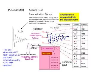
Generation and Fourier Transformation (FT) of a Free Induction Decay (FID) Pattern Complex summation wave (FID) Four different frequencies Fourier transformation http://www.cem.msu.edu/~reusch/VirtualText/Spectrpy/nmr/nmr2.htm#pulse
Free Induction Decay (FID) Signal: A Decaying Cosine Curve 5 Hz signal Assume T2 = 2 sec It = Ioe-t/T2 ~35% of signal remains after 2 sec. 0 1 2 3 4 5 seconds http://www.cem.msu.edu/~reusch/VirtualText/Spectrpy/nmr/nmr2.htm#pulse
Portion of the FID of Betulin 0.02 0.04 0.06 0.08 0.1 Time (sec)
= chemical shift (Hz) – shift of tetramethylsilane (TMS; 0 Hz) = ppm spectrometer frequency (MHz) Increasing deshielding Downfield Upfield Increasing shielding, Bo These effects are cumulative, so the presence of more electronegative groups produce more deshielding and therefore, larger chemical shifts.
In a magnetic field, the six electrons in benzene circulate around the ring creating a ring current. • The magnetic field induced by these moving electrons reinforces the applied magnetic field in the vicinity of the protons. • The protons thus feel a stronger magnetic field and a higher frequency is needed for resonance. Thus they are deshielded and absorb downfield.
If the formula is known ( C8H9OF ), add up all of the “steps” and divide by the number of hydrogens = (24 + 16 + 32 mm) / 9H = 8.0 mm / Hydrogen. a = 24 mm / 8.0 mm/H 3 H; b = 16 mm/8.0 mm/H 2H; c = 32 mm/8.0 mm/H 4H.
Splitting pattern: how many neighboring hydrogens. • In general, n-equivalent neighboring hydrogens will split a 1H signal into an ( n + 1 ) Pascal pattern. • “neighboring” – no more than three bonds away • n n + 1 Pascal pattern: • 0 1 1 singlet • 1 2 1 1 doublet • 2 3 1 2 1 triplet • 3 4 1 3 3 1 quartet • 4 5 1 4 6 4 1 quintet
note: n must be equivalent neighboring hydrogens to give rise to a Pascal splitting pattern. If the neighbors are not equivalent, then you will see a complex pattern (aka complex multiplet). note: the alcohol hydrogen –OH usually does not split neighboring hydrogen signals nor is it split. Normally a singlet of integration 1 between 1 – 5.5 ppm (variable).
Nuclear Magnetic Resonance Spectroscopy 13C NMR • The lack of splitting in a 13C spectrum is a consequence of the low natural abundance of 13C. • Recall that splitting occurs when two NMR active nuclei—like two protons—are close to each other. Because of the low natural abundance of 13C nuclei (1.1%), the chance of two 13C nuclei being bonded to each other is very small (0.01%), and so no carbon-carbon splitting is observed. • A 13C NMR signal can also be split by nearby protons. This 1H-13C splitting is usually eliminated from the spectrum by using an instrumental technique that decouples the proton-carbon interactions, so that every peak in a 13C NMR spectrum appears as a singlet. • The two features of a 13C NMR spectrum that provide the most structural information are the number of signals observed and the chemical shifts of those signals.
Nuclear Magnetic Resonance Spectroscopy 13C NMR—Number of Signals • The number of signals in a 13C spectrum gives the number of different types of carbon atoms in a molecule. • Because 13C NMR signals are not split, the number of signals equals the number of lines in the 13C spectrum. • In contrast to the 1H NMR situation, peak intensity is not proportional to the number of absorbing carbons, so 13C NMR signals are not integrated.
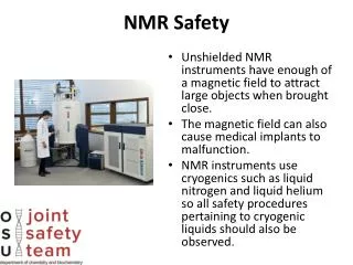
MRI • Magnetic resonance imaging, noninvasive • “Nuclear” is omitted because of public’s fear that it would be radioactive. • Only protons in one plane can be in resonance at one time. • Computer puts together “slices” to get 3D. • Tumors readily detected. => Chapter 13