Download
1 / 35
350 likes | 620 Views
Male genital system. Histology and embryology. Development. Development of gonads Development of genital ducts Development of external genital. Origin. Gonads – intermedial mesoderm of mesonephros Primordial germ cells – endoderm of yolk sac External genitalia – ectoderm and mesoderm.

E N D
Male genital system Histology and embryology
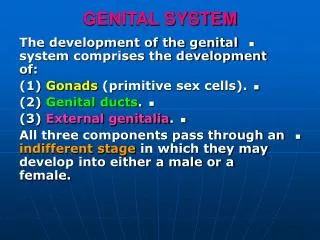
Development • Development of gonads • Development of genital ducts • Development of external genital
Origin • Gonads – intermedial mesoderm of mesonephros • Primordial germ cells – endoderm of yolk sac • External genitalia – ectoderm and mesoderm
Indiferent stage • Both sexes has same first stage • coelomic epithelium • primary germ cords • primordial germ cells • mesonephric duct (Wollfian) and tubules • paramesonephric duct
Male gonads • Y – chromosome • SRY -> TDF (testes determining factor) • if produced -> development of testis • usually from 7th week • if not produced -> development of ovarium • usually from 12th week • „waiting period“
Development of testis • TDF stimulates proliferation of primary germ cord -> medullary cords • origin of seminiferous tubules and rete testis • origin of Sertoli cells • intermedial mesenchyme • origin of Leydig cells • rest of coelomic epithelium changed to tunica albuginea
Seminiferous tubules • spermatogonia – from primordial germ cells • Sertoli cells – surrounds spermatogonia • secrete AMH – anti-müllerian hormone • inhibition of paramesonephric duct (Müllerian) • interstitial Leydig cells • produce testosteron from 8th week • no lumen till puberty
Descensus of the testis • from thoracolumbal area to scrotum • AMH, testicular growth, elongation of body • gubernaculum – connective cord between testis and scrotum • from 26th week • mark of fetal maturity • cryptorchism vs. ectopic testis
Genital ducts • Connected medullar cords • rete testis • Mesonephric tubules • Efferent ducts • Mesonephric duct (Wollfian) • Epididymal duct, ductus deferens, vesicular glands, ductus ejaculatorius • (ureter, pelvis, calices, collecting duct and tubules) • Paramesonephric duct (Müllerian) disappear
indiferent stage genital tubercle urogenital folds labioscrotal folds male genital penis spongious urethra scrotum External genitalia
Histology Testis Ducts Accesory glands
Testis • Tunica albuginea • dense connective tissue - collagenous • Lobuli testis • Seminiferous tubules • Interstitium
Seminiferous tubules • Stages of spermatogonia surrounded by Sertoli cells • hemato-testicular barrier – occluding junctions between medial part of Sertoli cells • nutrition, support • earlier stages are on the periphery of tubule • production of AMH, inhibin • regulated by follicle stimulating hormone
Spermatogenesis • development of sperm from spermatogonia • multiple division – two times meiotic • spermiogenesis – maturation of spermatid into sperm
Sperm • maturation is 64 days long (from first division of spermatogonia A) • Head • Acrosome (enzymes), nucleus • Connecting piece - pair of centrioles • Middle piece • mitochondrial sheet, outer dense fibers (9), axonema • Principal piece • ribs of the fibrous sheet, outer dense fibers (7), axonema
Testicular interstitium • Blood vessels • Lymphatic vessel • Leydig cells • production of testosteron • stimulation of spermatogenesis, maintanance of sex glands, sebaceous glands ... • regulated by luteinizing hormon of hypophisis
Sperm maturity and transport pathway • Tubuli recti et rete testis • Epididymis • Ductus deferens • Ejaculatory duct
Tubuli recti et rete testis • cuboideal Sertoliho cells • no spermatogenic cells • surrounded by lymphatic channels, blood vessels and Leydig cells
Epididymis • Ductuli efferentes - in head • columnar epitheluim with stereocilia and cilia • non-motile sperm • scalloped outline • smooth muscle layer
Epididymis • Ductus epididymidis - in body & tail • highly coiled one tubule - 4-6 m long • pseudostratified columnar epi with stereocilia • two layers of smooth muscle - thicker • function • maturation • storage • transport
Spermatic cord • Vessels, muscles, lymphatic vessels, nerves • Ductus defernes (Vas deferens) • pseudostratified columnar epi • lamina propria • 3 layers of smooth muscle (L, C, L)
Ejaculatory duct + Ampulla • pseudostratified columnar epi • lamina propria • muscular layer only in ampulla
Accesory Genital glands • Seminal vesicles • Prostate • Bulbourethral glands
Seminal vesicles (Glandulae vesiculosae) • cuboidela-to-pseudostratified epi • folded mucosa • smooth muscle layer (C+L) • adventitia + serosa on apex ! • function: 50-70% of seminal fluid • fructose
Prostate • 30-50 tuboalveolar glands in fibromuscular stroma • 3 regions of glands • periurethral mucosal gl. • periurethral submucosal gl. • peripheral main gl.
Prostate • simple-to-pseudostratified epi • prostatic concrements (corpora amylacea) • protein rich material, calcifications • function: enrich the semen fluid, fibrinolysin, amylase, PSA
Bulbourethral glands • mucus secreting glands • +galactose • lubrication function
Penis • three masses of erectil tissue • 2x corpora cavernosa • 1x corpus spongiosum • communicating blood spaces (sinuses) surrounded by connective tissue • artery -> sinus -> vein • dilatation of artery (NO) compress vein, sinuses fill with blood -> erection occurs