Download
1 / 114
1.18k likes | 1.36k Views
The Cardiovascular System: Blood. Chapter 14. Function. Transportation-hormones, gasses, nutrients, ions, heat Regulation- pH, temperature, water balance in cells Protection- clotting, white cells interferons, complement. Composition. Connective tissue-Two parts

E N D
The Cardiovascular System: Blood Chapter 14
Function • Transportation-hormones, gasses, nutrients, ions, heat • Regulation- pH, temperature, water balance in cells • Protection- clotting, white cells interferons, complement
Composition • Connective tissue-Two parts • Plasma = soluble materials (~55%) • Formed Elements = cells (~45%) • Percent occupied by red blood cells (RBC) = hematocrit (Hct) • White blood cells (WBC) ~1%
Plasma • ~91% water, 7% proteins, 1.5 % other solutes • Proteins: Albumin (54%)- osmosis and carriers; • Globulins (38%)- antibodies • Fibrinogen (7%)- clotting • Other: Electrolytes , nutrients, gases, hormones, vitamins & waste products
Formed Elements I. Red Blood Cells II. White blood cells • A. granular Leukocytes • Neutrophils • Eosinophils • Basophils • B. Agranular leukocytes • T & B lymphocytes & natural Killer cells • monocytes III Platelets
Formation of Blood Cells • Called hemopoiesis • Just before birth and throughout life occurs in red bone marrow • Contains pluripotent stem cells • In response to specific hormones these develop through a series of changes to form all of the blood cells
Erythrocytes (RBCs) • Hemoglobin package- carries oxygen • Also carries some CO2 • Male has ~ 5.4 million cells/µl; Female has ~4.8 million • membrane, no nucleus, flexible structure • use glucose for ATP production to maintain ionic composition • No mitochondria • Wear out fast- live ~120 days
RBC Cycling • cleared by macrophages (liver & Spleen) • Fe- recycled in bone marrow • Carried in blood on transferrin • Heme bilirubin and excreted (bile) • Globin A.A. recycled.
RBC Synthesis • called erythropoiesis • From stem cells: hemocytoblasts • Released as reticulocytes • Mature to erythrocytes in 1-2 days • Production & destruction is balanced • Low O2 delivery (hypoxia) • erythropoietin release (EPO) fromkidney • Stimulates erythropoiesis
White Blood Cells • Defenses: phagocytes, antibody production and antibacterial action • Phagocytes: • Neutrophil- first responders • Monocytes macrophages (big eaters) • Eosinophil- phagocitize antibody-antigen complexes Involved in suppressing allergic responses • Basophil- intensify allergic reactions • Immune response: • T-cells, B-cells& natural killer (NK) cells
WBC Life Span • 5000-10,00 WBC /µl blood • Limited number of bacteria can be eaten • Life span is a few days • During active infection may be hours • Leukocytosis= increased WBC numbers response to stresses • Leukopenia = decreased WBC numbers
Platelets • Myeloid stem cells megakaryocytes 2000 -3000 fragments = platelets • Plug damaged blood vessels • Promote blood clotting • Life span 5-9 days
Hemostasis • Hemostasis = stationary blood • 1. Vascular reactions (spasm) • Response to damage • Quick reduction of blood loss • 2. platelet plug formation • Become sticky when contact damaged vessel wall • 3. blood clotting (coagulation) • Series of chemical reactions involving clotting factors • Clotting in unbroken vessel=thrombosis
Coagulation • Extrinsic pathway common steps • tissue factor(TF) from damaged cells 1 • Intrinsic Pathway common steps • Materials “intrinsic” to blood 1 • 1. prothrombinase which causes • 2. prothrombin thrombin causes • 3. fibrinogen fibrin clot
Clot Retraction & Vessel Repair • Clot pugs ruptured area • Gradually contracts (retraction) • Pulls sides of wound together • Fibroblasts replace connective tissue • epithelial cells repair lining
Control Mechanisms • Fibrinolysis: dissolving of clot by activated plasmin enclosed in clot • Clots can be triggered by roughness on vessel wall = thrombosis • Loose clot = embolus and can block a small vessel = embolism
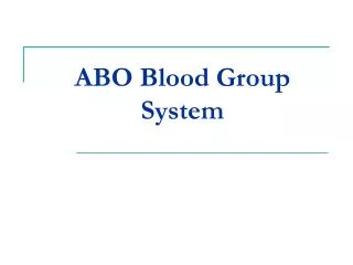
Blood Types • Surface antigens- react with antibodies • Divided into groups based on antigens • > 24 blood groups and > 100 different antigens • We will deal with ABO and Rh groups
ABO Group • Two antigens = A & B • If have only A –type A • If have only B –type B • If neither then Type O • Blood usually has antibodies that can react with antigens • e.g. anti-A antibody or anti-B antibody • You don’t react with your own antigens • Thus: type A has anti-B and vice versa
Rh Blood Group • Antigen discovered in rhesus monkey • If have antigen- Rh+ • Normally don’t have antibodies • antibodies develop after the first exposure from transfusion
Transfusions • If mismatched blood given antibodies bind to it and hemolyze cells • Type AB has no AB antibodies so can receive any ABO type blood called Universal recipients • Type O have neither antigen so can donate to any other ABO type called Universal donors • Misleading because of many other blood groups that must be matched
The Cardiovascular system: Heart Chapter 15
Location • Thoracic cavity between two lungs • ~2/3 to left of midline • surrounded by pericardium: • Fibrous pericardium- • Inelastic and anchors heart in place • Inside is serous pericardium- double layer around heart • Parietal layer fused to fibrous pericardium • Inner visceral layer adheres tightly to heart • Filled with pericardial fluid- reduces friction during beat.
Heart Wall • Epicardium- outer layer • Myocardium- cardiac muscle • Two separate networks via gap junctions in intercalated discs- atrial & ventricular • Networks- contract as a unit • Endocardium- Squamous epithelium • lines inside of myocardium
Chambers • 4 chambers • 2 upper chambers= Atria • Between is interatrial septum • Contains fossa ovalis- remnant of foramen ovalis • 2 lower chambers = ventricles • Between is interventricular septum • Wall thickness depends on work load • Atria thinnest • Right ventricle pumps to lungs & thinner than left
Great Vessels Of Heart- Right • Superior & inferior Vena Cavae • Delivers deoxygenated blood to R. atrium from body • Coronary sinus drains heart muscle veins • R. Atrium R. Ventricle • pumps through Pulmonary Trunk • R & L pulmonary arteries • lungs
Great Vessels Of Heart-Left • Pulmonary Veins from lungs • oxygenated blood • L. atrium Left ventricle • ascending aorta body • Between pulmonary trunk & aortic arch is ligamentum arteriosum • fetal ductus arteriosum remnant
Valves • Designed to prevent back flow in response to pressure changes • Atrioventricular (AV) valves • Between atria and ventricles • Right = tricuspid valve (3 cusps) • Left = bicuspid or mitral valve • Semilunar valves near origin of aorta & pulmonary trunk • Aortic & pulmonary valves respectively
Blood Supply Of Heart • Blood flow through vessels in myocardium = coronary circulation • L. & Right coronary arteries • branch from aorta • branch to carry blood throughout muscle • Deoxygenated blood collected by Coronary Sinus (posterior) • Empties into R. Atrium
Conduction System • 1% of cardiac muscle generate action potentials= Pacemaker & Conduction system • Normally begins at sinoatrial (SA) node • Atria & atria contract • AV node -slows • AV bundle (Bundle of His) • bundle branches Purkinje fibers • apex and up- then ventricles contract
Pacemaker • Depolarize spontaneously • sinoatrial node ~100times /min • also AV node ~40-60 times/min • in ventricle ~20-35 /min • Fastest one run runs the heart = pacemaker • Normally the sinoatrial node