Download
1 / 42
420 likes | 517 Views
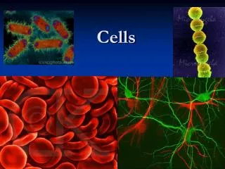
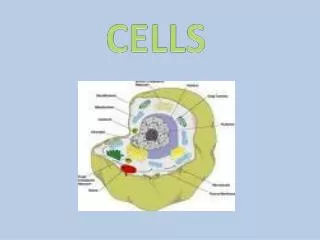
Cells. Chapter 3. Generalized View of the Cell. There are three main parts to a cell and each part has a very specific function. Read pages 49-50 to discover them on your own…. The Plasma Membrane. Cytoplasm.

E N D
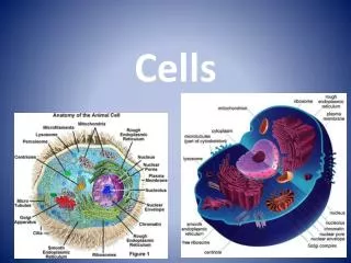
Cells Chapter 3
Generalized View of the Cell • There are three main parts to a cell and each part has a very specific function. • Read pages 49-50 to discover them on your own….
Cytoplasm • Cytoplasm consists of fluid inside plasma membrane and membrane-bound organelles except the nucleus • Cytosol consists of fluid inside plasma membrane but excludes nucleus and membrane-bound organelles
Transport Across the Plasma Membrane • Fluids in average body = ~60% • ICF -- inside the cell (cytosol) • ECF – outside the cell • Interstitial fluid (between cells of tissues) • Plasma (blood vessels) • Lymph (lymphatic vessels) • Cerebrospinal fluid (within and around brain/spinal cord) Materials dissolve into these body fluids; direction of movement dependent upon concentration (amount of solute in solution)
Concentration Gradients • Differences between ICF and ECF in solute concentration 3% salt solution 5% salt solution ??? Water ??? Water PassiveActive High to low Low to high No energy needed Energy needed
Concentration Gradients • Differences between ICF and ECF in solute concentration 3% salt solution 5% salt solution 97% Water 95% Water Passive High to low until dynamic equilibrium reached Down concentration gradient No energy needed
Passive Processes • Does not require the use of energy • Diffusion defined: • Substance moves due to kinetic energy • Movement from high concentration to low concentration • Movement of more molecules in one direction is called net diffusion • Movement ‘down the concentration gradient’ • Continues until equilibrium is reached
Two types of diffusion • Simple diffusion: lipid-soluble substances, simple cross membrane down the gradient • Facilitated diffusion: ions, through pores of ion channels of integral proteins
Osmosis • Net movement of water down the gradient; lower solute concentration to higher solute concentration through • Lipid bilayer • Integral proteins 20% sucrose 80% water
Osmotic Pressure • Pressure exerted on plasma membrane due to a solution containing solute particles that cannot pass through membrane • Higher solute concentration = higher osmotic pressure • Lower solute concentration = lower osmotic pressure
Osmotic Solutions • Isotonic solution: cells maintain normal shape and volume; concentration of solutes equal on both sides of membrane • Hypotonic solution: higher concentration of water outside; higher concentration of solutes than cytosol inside cell • Water molecules will enter cell faster than they leave it = cell will swell, eventually burst • Bursting of red blood cells referred to as hemolysis • Hypertonic solution: higher concentration of water inside; lower concentration of solutes than cytosol inside cell • Water molecules will leave cell faster than they enter it = cell will shrink • Shrinkage of red blood cells referred to as crenation
Short Video Review http://www.hartnell.edu/tutorials/biology/osmosis.html
Passive or Active? • Have I been talking about passive, active, or passive and active transport?
Active Transport • From low to high concentration; ‘up the concentration gradient’ • Requires the use of energy • Comes from splitting of ATP molecule • Changes shape of transporter protein, called a pump • Transports ions: Na+, K+, H+, Ca+2, I-, Cl- • Example: sodium-potassium pump • 40% of a cell’s ATP expended on active transport • Drugs like cyanide can turn off ATP production--FATAL
Cyanide • Cyanide can be a colorless gas, such as hydrogen cyanide (HCN) or cyanogen chloride (CNCl), or a crystal form such as sodium cyanide (NaCN) or potassium cyanide (KCN). • Cyanide sometimes is described as having a “bitter almond” smell, but it does not always give off an odor, and not everyone can detect this odor. • You could be exposed to cyanide by breathing air, drinking water, eating food, or touching soil that contains cyanide. • Cyanide enters water, soil, or air as a result of both natural processes and industrial activities. When present in air, it is usually in the form of gaseous hydrogen cyanide. • Smoking cigarettes is probably one of the major sources of cyanide exposure for people who do not work in cyanide-related industries.
Transport in Vesicles • Vesicles small sacs formed by budding off of membranes • Transport substances within the cell from one structure to another • Energy source again is ATP • Take in substances from ECF and transport substances out to ECF • Endocytosis: materials moved into cell • Phagocytosis ‘to eat’ - solids • Bulk-phase endocytosis(pinocytosis) liquids • Exocytosis: materials moved out of cell
Endocytosis • Endocytosis: capturing substance or particle from outside the cell by engulfing it within membrane folds from the cell membrane and releasing it into cytosol. • There are two main kinds of endocytosis: • Phagocytosis • Bulk-phase endocytosis (pinocytosis)
Phagocytosis • Phagocytosis ”cellular eating” • Particles bind to plasma membrane receptors • Projections called pseudopods extend surround particles and portions of the membrane fuse to form a vesicle • Extensions of the plasma membrane and cytoplasm • Pseudopods vesicle formed called a phagosome • Phagosome enters the cell, fuses with lysosomes • Lysosome enzymes break down phagosome’s contents • Any undigested content remains in the phagosome, now called a residual body Occurs only in phagocytes (certain white blood cells and macrophages), cells specialized to engulf and destroy bacteria, viruses, aged dying cells, and foreign matters protecting body from disease
Bulk-phase endocytosis (pinocytosis) • Bulk-phase endocytosis (pinocytosis) ”cellular drinking” • Plasma membrane folds inward, forming a vesicle allowing tiny droplets of extracellular fluid that contain dissolved substances to be surrounded • Vesicle detaches or “pinches off” of the plasma membrane and enters the cytosol • Liquid is encircled within a pinocytic vesicle • Vesicle fuses with a lysosome, enzymes degrade engulfed solutes • Degraded solutes; like amino acids and fatty acids leave the lysosome to be used elsewhere in the cell
Exocytosis • Exocytosis: process of vesicles fusing with the plasma membrane and secretes their contents to the outside of the cell. • All cells do exocytosis process, but most important in: • Secretory cells • Release digestive enzymes, hormones, mucus, and other secretions • Nerve cells • Release neurotransmitters
Cytoplasm • Consists of all the cellular contents between the plasma membrane and the nucleus and includes both cytosol and organelles.
Cytosol • Cytosol is the liquid portion of the cytoplasm that surrounds the organelles and makes up about 55% of the cell’s volume. • 75%-90% of cytosol is water, the rest is composed of dissolved solutes and suspended particles. • Examples: ions, glucose, amino acids, fatty acids, proteins, lipids, ATP, and waste. • Site of many chemical reactions • Maintain cell structure and enable cell growth
Cytoskeleton • Extends throughout cytosol • Network of three different types of protein filaments: • Microfilaments • Intermediate filaments • Microtubules
Microfilaments • Contribute to cell strength and shape • Function: • Provide mechanical support and help generate movement • Anchor cytoskeleton to integral proteins • Provide support for microvilli Microvilli • Fingerlike projections of the plasma membrane • Increase cell surface area • Found mostly in areas with great absorption needs like the small intestines • Help cells attach to one another or extracellular materials • Involved in muscle contractions, cell division, and cell locomotion • Migration of embryonic cells • Invasion of tissues by white blood cells (WBCs) to fight disease • Migration of skin cells in wound healing
Intermediate Filaments & Microtubules • Intermediate Filaments • Found in parts of cells subjected to tension (stretching) • Hold organelles in place • Attach cells to one another • Microtubules • Long, hollow tubes • Help determine cell shape • Function as transport system for • Organelle movement • Secretory vesicles • Migration of chromosomes • Create movement of cilia and flagella
Organelles • Functions and identification of the organelles are your responsibility since this a total biology review area • Information found on pages 58-62 of your textbook
Lysosomes • Membrane-enclosed vesicles • May contain up to 60 different digestive enzymes • Fuse with other vesicles during endocytosis • Recycle the cell’s own structures (worn-out organelles) autophagy • May destroy own cell autolysis • This cause tissue deterioration after death • Faulty lysosomes can contribute to certain diseases, i.e. Tay-Sachs disease
Peroxisomes • Smaller than lysosomes • Contain enzymes called oxidases that oxide (remove hydrogen atoms from) various substances • Creating a by-product of hydrogen peroxide H2O2 • Potentially toxic compound associated with free radical superoxides • BUT peroxisomes also contain catalase which breaks down H2O2 • Oxidize toxic substances • Abundant in liver
Proteasomes • Tiny, barrel-like structure • Destroys unneeded, damaged, or faulty proteins from the cytosol • Contain enzyme called protease • Cuts proteins into small peptides • So other enzymes can break them down to amino acids from which new proteins can be built
Nucleus • On your own, this is also a biology review topic • Label the diagram on page 14 of your packet • List the functions of the nucleus also • Information found on page 62 of your textbook
Gene Action: Protein Synthesis • On your own, this is also a biology review topic • Information found on pages 64-65 of your textbook.
Somatic Cell Division Damaged, diseased, or worn out cells are replaced • Two types of cell division: • Reproductive cell division-meiosis • Will be discussed in later chapters • Somatic cell division-mitosis • Division into two identical cells Division occurs through a sequence of changes called the cell cycle • Two major parts to cell cycle • Interphase: when cell is not dividing • Mitotic phase: when cell is dividing
Interphase • 1st step is DNA replication • Then production of new organelles and cytoplasmic components fro the new cell • High metabolic activity • A lot of cell growth
Mitotic Phase Mitosis followed by Cytokinesis (splitting of the cytoplasm) Chromosomes are visible during this phase under a microscope
Nuclear Division: Mitosis Four stages: • Prophase • Chromatin condenses into visible chromosomes • Centrioles migrate to opposite poles • Mitotic spindles form attach to centromeres of chromatids • Nuclear envelop breaks down • Metaphase • Chromatids line up at equator (metaphase plate) • Anaphase • Centromeres split chromatids into chromosomes • Chromosomes dragged towards the poles • Telophase • Chromosomes uncoil into chromatin • Nuclear envelops reforms • Nucleolus reappear • Mitotic spindles break down
Cytoplasmic Division: Cytokinesis • Division of cytoplasm and organelles between two new cells • Begins with formation of a cleavage furrow in plasma membrane that pinches inward • Cells return to Interphase
Cellular Diversity • Average humans has about 100 trillion cells of varying sizes • Cell size is measured in micrometers (µm) • 1 micrometer = 1 one-millionth of a meter • Largest cell in human body is an oocyte with a diameter of 140 µm • Average hair strands is ~100 µm in diameter • Cells can be round, oval, flat, cube-shaped, column-shaped, elongated, star-shaped, cylindrical, or disc-shaped • Shape is related to function
Round: oocyte Oval: Liver Flat: Squamous Cube-shaped: Cubiodal Column-shaped: Collumnar Elongated: Collagen Star-shaped: Natural Killer Cells Cylindrical: Skeletal Disc-shaped: RBCs
Aging and Cells • As we age our cells ability to divide is diminished. • DNA sequences that code for cell division break down. • Free radical control becomes limited. • Autoimmune responses slow down.