Download

1 / 33
340 likes | 372 Views
Explore how the bilaminar embryonic disc transforms into a trilaminar structure during the second and third weeks of prenatal development, detailing the formation of amniotic cavity, primitive yolk sac, extraembryonic mesoderm, and more.

E N D
Bilaminar & Trilaminar Embryonic Disc Dr. Zeenat Zaidi
Bilaminar Embryonic Disc The Second Week
Formation of Amniotic Cavity As implantation of the blastocyst progresses, changes appear in the inner cell mass (embryoblast) A cavity, amniotic cavity appears separating embryoblast from the trophoblast, which soon becomes lined by amnioblasts derived from inner cell mass The cavity gradually increases in size and is filled with amniotic fluid
Formation of Embryonic Disc The inner cell mass becomes flattened forming a circular bilaminar plate, the embryonic disc, consisting of two layers: Epiblast (ectoderm), thicker layer, consists of high columnar cells, and is related to the amniotic cavity Hypoblast (endoderm),consists of small cuboidal cells, and lies adjacent to the blastocyst cavity
Formation of Primitive Yolk Sac The blastocyst cavity becomes lined with exocelomic membrane and is called exocelomic cavity The hypoblastic cells soon replace the exocelomic membrane and the cavity is then named as the primitive (primary) yolk sac
At this stage the embryonic disc is: A circular bilaminar disc, that lies between the amniotic cavity and the primitive yolk sac The epiblast forms the floor of the amniotic cavity & the hypoblast lies in the roof of the primitive yolk sac
Formation of Extraembryonic Mesoderm Endoderm of the yolk sac gives rise to a layer of loosely arranged connective tissue, extraembryonic mesoderm (EEM), which surrounds the amniotic cavity and the yolk sac.
Formation of Extraembryonic Celome Isolated spaces appear in the EEM These spaces rapidly fuse to form a large fluid filled, C-shaped cavity, the extraembyonic celome surrounding the amniotic cavity and the yolk sac
Formation of Connecting Stalk The region where no cavity has appeared, forms the connecting stalk, that connects the amniotic cavity, yolk sac and the embryonic disc to the outer wall The site of the connecting stalk determines the caudal pole of the embryonic disc
With the formation of extraembryonic celome: The extraembryonicmesoderm splits into two layers: an outer extraembryonic parietal (somatic)mesoderm an inner extraembryonicvisceral (splanchnic) mesoderm
The primary yolk sac decreases in size and becomes the secondary (definitive) yolk sac Wall of the yolk sac, amnion & chorion are formed
Amnion: Two layers: Amnioblasts Extraembronic splanchnic mesoderm Wall of the yolk sac: Two layers: Endoderm Extraembronic splanchnic mesoderm Chorion: Three layers: Extraembryonic somatic mesoderm Cytotrophoblast Syncytiotrophoblast
chorion amnion bilaminar disc wall of the yolk sac Connecting stalk EEC Extraembryonic celome is now called the CHORIONIC CAVITY
Trilaminar Embryonic Disc The Third Week The significant event of third week is Gastrulation
Gastrulation The process by which the bilaminar disc is converted into a trilaminar disc It is the beginning of morphogenesis (formation of body form) Consists of formation of the primitive streak, the threegerm layers & the notochord Embryo is referred to as a Gastrula
Primitive Streak The primitive streak results from proliferation of the epiblastic cells in the median plane, in the caudal half of the epiblast, and lies along the cranio-caudal axis. Its cranial end forms primitive node A groove, primitive groove, appears in the primitive streak, which continues with a small depression, primitive pit, in the primitive node.
A circular thickening appears in the hypoblast near the cranial end, in the midline, to form the prechordal plate, that marks the future site of mouth A circular thickening appears in the hypoblast caudal to primitive streak in the midline to form the cloacal membrane, the future site of the anus
By this stage of development, it is possible to identify the embryo’s: craniocaudal axis cranial and caudal ends dorsal and ventral surfaces right and left sides. Connecting stalk
Formation of Intraembryonic Mesoderm The epiblastic cells from the primitive streak (groove) proliferate to form mesenchymal tissue The newly formed cells invaginate, migrate ventrally, laterally & cranially between the epiblast and hypoblast & organize to form the intraembryonic mesoderm
Formation of Intraembryonic Mesoderm cont’d Intraembryonic mesoderm merges with the extra-embryonic mesoderm at the periphery of the embryonic disc By the end of 3rd week, mesoderm lies between embryonic ectoderm and endoderm everywhereexcept in the region of prechordal plate and cloacal membrane, as the embryonic ectoderm & endoderm are fused at these regions
Formation of Intraembryonic Mesoderm cont’d Some mesenchymal cells displace the hypoblasts forming the embryonic endoderm Cells remaining in the epiblast form the embryonic ectoderm
Each of the three germ layers gives rise to specific tissues and organs Thus the EPIBLAST gives rise to all three germ layers,Ectoderm, Mesoderm, Endoderm in the embryo
Fate of Primitive Streak Actively forms mesoderm until the early part of 4th week Then it starts regressing and becomes an insignificant structure in the sacrocooccygeal regions Normally it degenerates and disappears by the end of 4th week Remnants may persist and give rise to a large tumor called Sacrococcygeal Teratomas
Notochord A rod of mesenchymal cells located cranially, in the midline, extending between the primitive node and the prechordal plate
Formation of Notochord Mesenchymal cells migrate cranially from primitive pit toward the prechordal plate, and form a rod like notochordal process The notochordal process becomes canalized forming a hollow tube, the notochordal canal, communicating with the primitive pit.
Formation of Notochord cont’d The floor of the tube and the underlying endoderm break down, forming a notochordal plate The notochordal plate becomes continuous with the endodermal layer.
Formation of Notochord cont’d A temporary communication is established between the amniotic cavity and the yolk sac, termed the neurenteric canal.
Functions of Notochord Defines primordial axis of the embryo Provides rigidity to the embryo Serves as a basis for the development of the axial skeleton Indicates the future site of the vertebral bodies/column Regulates differentiation of surrounding structures including the overlying ectoderm (neural plate) and mesoderm (somites).
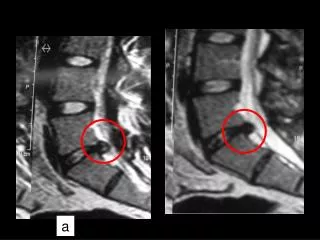
Fate of Notochord Degenerates and disappears as the bodies of the vertebrae develop, but it persists as the nucleus pulposus of each intervertebral disc Remnants of notochordal tissue give rise to tumors called Chordomas
Differentiation of the Intraembryonic Mesoderm Induced by the notochord Differentiates (in the region of notochord) into: Paraxial mesoderm Intermediate cell mass Lateral plate mesoderm