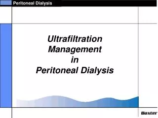
Ultrafiltration
Ultrafiltration. 1 Basic Ideas. Ultrafiltration is a effective separation method for proteins. Protein have two characteristics which are important for these separation. Large molecules Conformation change with pH. Osmotic Pressure.

Ultrafiltration
E N D
Presentation Transcript
1 Basic Ideas • Ultrafiltration is a effective separation method for proteins • Protein have two characteristics which are important for these separation • Large molecules • Conformation change with pH
Osmotic Pressure • Macromolecule is uncharged • Macromolecule can not pass through the membrane • Solvent flows from right to left, diluting the macromolecular sol’n • As the dilution takes place, the sol’n vol. increases and the level in the capillary rises Figure 1. Osmosis pressure across a membrane. Solvent can diffuse across the membrane shown, but solute cannot.
Ideal state If the macromolecular sol’n is dilute, then we can expand the logarithm in term of x1 Dilute sol’n : Van’t Hoff’s Law
Side chain of Proteins • Carboxylic acid ( - COOH ) : glutamic acid in basic sol’n to form carboxylate ( - COO- ) groups • Amine ( - NH2 ) : lysine in acid sol’n to form ammonium ( - NH3+ ) groups • A function of the pH of the sol’n : the relative amount of these positive and negative charges - Low pH : more -NH3+ and -COOH, High pH : more -NH2 and -COO- Figure 2. Charges on a protein. At low pH, amine side chains are protonated to give a positive charge ; at high pH, carboxylic side chain ionizeto give a negative charge. The intermediate pH of no net charge is called the isoelectric point
Table 1. Molecular weights and isoelectric pH values of Proteins
Transport Equations • Ultrafiltration : the species transported - solvent Chief force - pressure Ultrafiltration from membrane separations, and not from convectional filtration Solvent velocity ∝ force on solvent : Darcy’s Law Figure 3. Ultrafiltration from a pressure difference.
Basic Eq’n for Ultrafiltration If the membrane rejects all solutes, then σ= 1 . If the membrane passes both solvent and solute, then σ= 0
Chemical potential includes all forms of energy acting on the solute Reference energy for a single protein sphere Free energy of mixing If the sol’n is dilute, C is const. and the resulting term kBTln C is lumped into the μ10. The effect of gravitational force The solute charge and the electrical potential
0 1. Charged solute in the absence of any gravitatinal field 0 Gravitational force = 0 The solute charge = 0 : Fick’s Law
0 0 2. The concentration of an uncharged solute is uniform : Sedimentation coefficient, with dimensions of time
0 0 3. The concentration differences and gravitational forces are minor Table 2. Mobility Spectrum for Normal Human plasma Components : Engineering form
0 Example 1 Yeast Ultrafiltration Ultrafiltration of a well stirred suspension containing 0.1 vol% yeast suspension gives a flux of 36 gal/ft2 day under a pressure difference of 130 psi. (a) What is the value of Lp ? The yeast cells will have a very high molecular weight, so that their molar Concentration and the resulting osmotic pressure will be small. At the same time, this large size leads to high rejection, so σ= 1. As a result from (b) What is the water velosity through the membrane? To find the velocity through the membrane, we need only convert the units of the flux
Example 2 The Transport of Ovalbumin Imagine a solution of 25℃ containing 0.004g/cm3 of ovalbumin, a protein of molecular weight 45,000. The solution is buffered at a pH of 3.5 and has the viscosity close to water, 8.9×10³ g/cm sec. Under these conditions, the protein has a charge of +10, a diffusion coefficient of 7.8×10⁻⁷ cm2/sec, and a sedimentation coefficient of 3.5×10⁻¹³ sec. (a) Estimate the diameter of the protein. The easiest way to estimate the protein’s size is from the diffusion coefficient, as suggested by The protein’s diameter is about 20 times larger than that of a water molecule and about 200 times smaller than that of a bacterium.
(b) What are the flux and velocity when this protein diffuses from the solution across a 1 cm film into pure water? The flux is found by integrating across the film to find The average velocity can be estimated either by integrating or by dividing the flux by the average conc. The latter is easier :
(c) What is the protein’s velocity under the influence of gravity? The velocity in a gravitational field is found from where the acceleration is the that due to gravity : This velocity is much less than that due to diffusion. In fact, as we expect, the mixing due to diffusion (I.e., to Brownian motion) swamps any separation due to gravity. It is only in the ultracentrifuge, where the acceleration far exceeds that of gravity, where these effects become significant. In the words of our students, “moles are worth more than gravity.”
(d) What are the flux and velocity due to a force of 1 volt/cm? The velocity under an electric field can be calculated from In each second, the protein moves 100 diameters. The flux can be found by multiplying the velocity times the conc. The flux under an electric field is several hundred times that due to diffusion. Again according to our students, “volts are worth more than moles”
In passing, we can use the data given to calculate the mobility, as suggested by This value is highter than normal because of the large on the protein at this pH.
9. 2 Ultrafiltration Figure 3. Different types of ultrafiltration. These processes are most easily classified by the size of solutes being separated. The difference between them are dominated by differences in membranes used (After Lonsdale, J. Memb. Sci. 10 81 (1982)) • Ultrafiltration is a membrane process. • Such a process depends on the ability of a permeable membrane to differentiate between solutes of different size. • Three distinctive characteristics • Use a high cross flow • Dominated by the membrane • Depend on the membrane geometry in the actual equipment
Crossflow • Ultrafiltration almost always involves a large flow across the membrane surface, perpendicular to the flux through the membrane. • When solid particles are being ultrafiltered, the cross flow minimizes the development of a filter cake which would retard the process • When a marcromolecular sol’n is being ultrafiltered, the cross flow reduces marcromolecule accumulation near the membrane surface. concentration polarization • Membranes • Made by spreading a thin layer of organic sol’n on water, glass, or an inert support • Porosity : about 80% • Average pore size : 0.1㎛ ~ 1.0 ㎛ Figure 4. Ultrafiltration membranes - 1
Made by drawing warm, nonporous films of polymers like PP. • Porosity : 35% • Thickness : 0.003 cm Figure 5. Ultrafiltration membranes - 2 • Made by exposing nonporose pores films of mica or polycarbonate to α radiation. • Then etched away with HF acid sol’n • Lowest porosity : ~3% • Similar permeability to the other types Figure 6. Ultrafiltration membranes - 3
Equipment • It consists of alternate layers of membrane, Support screen, and distribution chambers for feed and permeate • It has the smallest area per Vol. of the common types, and so tends to give low ultrafiltration fluxes per Vol. Figure 7. Membrane geometries for Ultrafiltration - Flat Sheets • The feed stream enters the lumen of the tubes, the permeate passes through the walls, and the retentate passes out other end of the tubes • It is harder to clean and service than the plate and flat sheet and it has lower area per Vol. and hence lwer fluxes than the spiral wound and hollow fiber geometries Figure 8. Membrane geometries for Ultrafiltration - Shell and Tube
This device is like a huge envelop made of membrane and containing a feed spacers • The device give higher filtration rates per Vol. • They are much harder to clean, and often must be discarded if even part of the membrane fails • They tend to be used when the feed is relatively pure, as in the production of ultrapure water by reverse osmosis Figure 9. Membrane geometries for Ultrafiltration - Spiral Wound • The fibers are typically 0.01 cm on diameter, while the tubes are around 1 cm diameter • Which configuration is best depends on the specific situation, but os rarely obvious Figure 10. Membrane geometries for Ultrafiltration - Hollow Fiber
Analysis • To find this time, we first must find the solvent velocity through the membrane This velocity is given by If the solute is completely rejected by the membrane, the reflection coefficient σ = 1 ; if the sol’n is dilute, the osmotic press. ΔΠ = RTc10, where c10 is the solute conc. at the surface of the membrane • Higher solute concentration Z = 0, c1 = c10 Z = l, c1 = c1 Boundary condition Integrating Figure 11. Conc. polarization.
A plot of flux versus the logarithm of reservoir conc. c1 should be a straight line Figure 12. Flux versus reservoirr conc. These data, for the ultrafiltration of B. Thuringlensis, support the analysis which leads to • Estimating the time to filter a given volume In this case , c10 = c1 Because the membrane rejects all the solute, the (n1 = c1V)is const.
Initial condition t =0, V = V0 Integrating
Example 2 The Surface Conc. For Chymotrypsin Ultrafiltration We are carrying the ultrafiltration of chymotrysin in a spiral wound module at a rate of 1.3×10⁻³ cm / sec (28 gal / ft2 day). The sol’n conc. is 0.44 wt%, the protein’s diffusion coefficient is 9.5×10⁻⁷ cm2 / sec, and the boundary layer is about 180×10-4 cm thick. How high is the surface conc. ? The conc. at the wall is 30% higher than that in the bulk
Example 3 Estimating the Time for Vaccine Ultrafiltration We want to ultrafilter 840 liters of a solution containing 0.061 wt% of a protein used as a vaccine for herpes. The protein has a diffusion coefficient of 1.1×10⁻⁶ cm2/sec and a molecular weight of 16,900. We would like to get the conc. up to about 2% by weight. The ultrafilter which we hope to use has eight hollow fiber cartridge, each of which has a surface area of 1.20 m2. It is cooled to 4℃. The membrane in these cartridges gives an pressure drop of 31 atm. (a) Assuming negligible conc. polarization, estimate the time to complete this filtration Since this is much less than the volume being filtered, even at the end of the filtration, we neglect this term 0
This ultrafiltration will take slightly less than two days-if conc. polarization remains unimportant. (b) Test whether conc. polarization is significant To see if conc. polarization is unimportant, we turn to to find This is greater than one, but not much greater than one. We would expect at least some effect of conc. polarization and would certainly make a laboratory test before risking all 840 liter of feed