
Enhanced Binding of APAP Moiety to p53 Protein in C6 Glioma Cells Post-APAP Exposure
This study investigates the binding of the acetaminophen (APAP) moiety to the p53 protein in C6 glioma cells. Cells were treated with DMSO or 5 mM APAP for 24 hours. Following treatment, equal amounts of the soluble protein fraction were used to immunoprecipitate p53 utilizing a specific antibody. The resulting p53 protein was analyzed via SDS-PAGE and immunoblotting with an antibody against APAP. Results indicate a significant increase in APAP binding to p53 after APAP exposure, highlighting the interaction between APAP and this critical tumor suppressor protein.

Enhanced Binding of APAP Moiety to p53 Protein in C6 Glioma Cells Post-APAP Exposure
E N D
Presentation Transcript
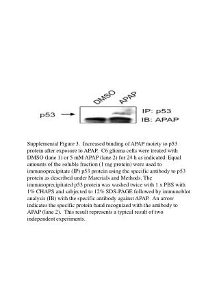
Supplemental Figure 3. Increased binding of APAP moiety to p53 protein after exposure to APAP. C6 glioma cells were treated with DMSO (lane 1) or 5 mM APAP (lane 2) for 24 h as indicated. Equal amounts of the soluble fraction (1 mg protein) were used to immunoprecipitate (IP) p53 protein using the specific antibody to p53 protein as described under Materials and Methods. The immunoprecipitated p53 protein was washed twice with 1 x PBS with 1% CHAPS and subjected to 12% SDS-PAGE followed by immunoblot analysis (IB) with the specific antibody against APAP. An arrow indicates the specific protein band recognized with the antibody to APAP (lane 2). This result represents a typical result of two independent experiments.