Download
1 / 79
830 likes | 1.23k Views
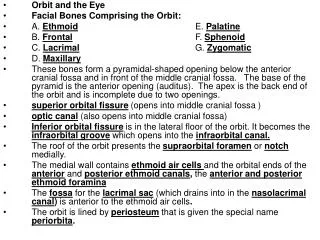
Bones of the Orbit. Protection, Grrr!. The Orbit. The orbits are bony cavities on either side of the midsagittal plane of the skull below the cranium that house the globe and orbital contents

E N D
Bones of the Orbit Protection, Grrr!
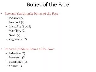
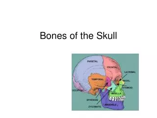
The Orbit • The orbits are bony cavities on either side of the midsagittal plane of the skull below the cranium that house the globe and orbital contents • They contain the globes, the extraocular muscles, and orbital nerves, blood vessels, and connective tissue for the proper functioning of the eye—all remaining space is filled with orbital fat • In addition, the orbits contain some vessels and nerves of passage, which serve areas of the face around the orbits • Each orbit is composed of 7 bones, since 3 of these bones (frontal, ethmoid, and sphenoid) are bones common to both orbits, 11 different bones are found in the two orbits (4 pairs and 3 common)
The Orbit 2 • The orbit is roughly the shape of a four-sided (quadrilateral) pyramid, the base of which is at the anterior orbital margin and the apex at the posterior orbital margin • Actually, the floor of the orbit is the shortest orbital wall and does not reach the apex, therefore, the orbit is more triangular in its posterior portion • The orbit develops around the eyeball and the lacrimal gland, so it has its greatest dimensions 1.5 cm behind the orbital margin • Whitnall described the orbit as pear shaped, with the stalk being the optic canal
The Orbit 3 • The orbital walls are referred to as the roof, floor, medial and lateral walls • The medial walls run approximately parallel to one another, whereas the lateral walls, if extended posteriorly, would form about a ninety-degree angle with one another • The following are the approximate dimensions of the orbit: • Depth = 4.5 cm • Width = 4 cm • Height = 3.5 cm
The Orbit 4 • The orbits are oriented forwards, outwards, and slightly downward • The angle between the lateral and medial wall of each orbit is about 45 degrees • The angle between the vertical planes of the two orbital entrances (the nasion angle) = 145 degrees • The orbital axis diverges about 22.5 degrees from the visual axis • New parents are sometimes concerned that their baby has a squint, however, this can be a false perception due to the more lateral displacements of the orbits and the smaller inter-orbital distance, since the bridge of the nose has not developed yet
Orbital Roof • The roof is triangular in shape and slopes downwards from the apex—it is concave anteriorly, but relatively flat posteriorly • It is formed by: • Orbital plate of the frontal bone • Lesser wing of the sphenoid posteriorly • The lateral border is marked by the zygomatico-frontal suture • The medial border of the roof is marked by the articulation of the frontal bone with the ethmoid, lacrimal, and maxilla bones • The roof of the orbit is fragile, thin, and somewhat transparent in the area separating the orbit from the anterior cranial fossa • In an elderly adult, bone in this area may be absorbed, leaving the orbital periosteal connective tissue (periorbita) in contact with the dura covering of the frontal lobe of the brain
Temporal Medial
Orbital Roof 2 • Landmarks: • Fossa for the lacrimal gland • Increased concavity at the antero-lateral border behind the zygomatic process of the frontal bone • Lodges the lacrimal gland anteriorly • Posteriorly, it contains orbital fat • Usually smooth, but may be pitted by the attachment of the suspensory ligament of the lacrimal gland • Trochlear fossa – small pit in the antero-medial portion of the roof, marking the attachment of the cartilaginous pulley of the superior oblique
Trochlear fossa Fossa for the lacrimal gland
Orbital Roof 3 • Usually the roof is smooth, but can show small apertures called the cribra orbitalia created by vessels during embryological development • The optic foramen marks the orbital opening of the optic canal, which projects posteriorly into the middle cranial fossa • The optic canal is formed by the two roots of the lesser wing of the sphenoid bone • The optic canal projects medially and posteriorly, forming an angle of 36 degrees to the median sagittal plane • The orbital opening of the canal is 6-6.5 mm high vertically and 4-4.5 mm wide horizontally—in the middle portion it is 5 x 5 mm
Orbital Floor • The floor is also triangular and not quite horizontal, but slopes slightly down from the medial to the lateral side • It is formed by: • Maxilla—large central area • Zygomatic—antero-lateral portion • Palatine—small posterior area • The floor is separated from the medial wall only by the fine suture formed by the articulation of the maxilla with the ethmoid bone
Orbital Floor 2 • It is separated from the lateral wall by: • Posteriorly, the inferior orbital fissure (sphenomaxillary fissure) • Anteriorly, it is continuous with the lateral wall, since the zygomatic bone forms both • Landmarks: • The infraorbital canal, running from the infraorbital fissure, buries deep into the maxilla and opens onto the face, about 4 mm below the orbital margin—the opening is the infraorbital foramen
Orbital Floor 3 • Landmarks (continued) • The infraorbital sulcus and canal transmit the infraorbital vessels and nerve—it also gives off the middle and superior alveolar (dental) canals for the corresponding nerves and vessels • Below the thin floor of the orbit, for nearly its entire extent is the maxillary sinus—therefore, tumors of that sinus can easily invade the orbit causing proptosis (the eye to protrude forward) • The lower orbital margin is made up of the maxilla and the maxillary process of the zygomatic bone
Blow-Out Fracture • A blow to the orbital rim can cause compression of the orbital contents, and such a sudden increase in intraorbital pressure might cause a fracture in one of the orbital walls. • In a classic blow-out fracture, the orbital rim remains intact. • The floor of the orbit is particularly susceptible to such a fracture, which usually occurs in the thin region along the infraorbital canal
Blow-Out Fracture 2 • Clinical signs and symptoms include: • Orbital swelling • Ecchymosis—blood escaping into tissue from damaged vessels • Anesthesia of the area served by the infraorbital nerve • Diplopia caused by restriction of the movement of the eye (particularly noted in upward gaze) • Limitations in ocular motility are caused by damage to the inferior extraocular muscles, either from bruising or hematoma, or from entrapment of the muscle or its associated connective tissue within the fracture
Medial Wall of the Orbit • The medial wall is the only one that is not triangular—it is oblong, either flat or convex toward the orbital cavity • It runs parallel to the median sagittal plane • It is formed by: • Maxilla bone • Lacrimal bone • Ethmoid—lamina papyracea • Body of the sphenoid—small portion
Medial Wall of the Orbit 2 • Landmarks: • Fossa for the lacrimal sac—anterior part of wall • Anterior lacrimal crest—frontal process of maxilla • Posterior lacrimal crest—lacrimal bone • Lacrimal tubercle—small thickening of bone at the lower part of the anterior lacrimal crest • Anterior and posterior ethmoidal foramina—in medial wall near the roof, in the frontoethmoidal suture
Medial Wall of the Orbit 3 • Landmarks (continued) • Thinnest of all orbital walls—translucent, ethmoidal air sinuses can be seen • Lamina papyracea (orbital surface of the ethmoid), as the name indicates, is thin as paper—infection easily spreads through it • Ethmoiditis is the most common cause of orbital cellulitis • The lacrimal bone is frequently absorbed during life
Lateral Wall of the Orbit • The lateral wall is triangular with the base anterior—it is slightly convex posteriorly, flat over the center, and concave anteriorly • It is formed by: • Greater wing of the sphenoid, posteriorly • Zygomatic bone, anteriorly • The sphenoidal portion is demarcated from the roof by the superior orbital fissure and from the floor by the inferior orbital fissure • The zygomatic portion is demarcated from the roof by its articulation with the frontal bone, but continues into the floor
Lateral Wall of the Orbit 2 • Landmarks: • Spina recti lateralis—a small bony projection on the inferior margin of the superior orbital fissure between the narrow and wide portions of the fissue • Gives origin to a part of the lateral rectus muscle • Produced mainly by a groove formed by the superior ophthalmic vein • Zygomatic groove and foramen • Groove runs from the anterior end of the inferior orbital fissure and ends at the zygomatic foramen
Lateral Wall of the Orbit 3 • Landmarks (continued) • Lateral Orbital Tubercle (of Whitnall) • Slight elevation on the orbital surface of the zygomatic just within the orbital margin and about 11 mm below the articulation with the frontal bone • Gives attachment to: • Check ligament of the lateral rectus muscle • Suspensory ligament of the eyeball (ligament of Lockwood) • Aponeurosis of the levator palpebrae superioris muscle • Lateral palpebral ligament • The lateral wall is the strongest, especially at the orbital margin
Orbital Margins • The orbital margin is formed by the frontal bone, zygomatic bone, maxillary bone, and a small portion of the lacrimal bone • The average horizontal dimension of the orbit is 4 cm, the average vertical dimension is 3.5 cm, and the average depth is 4.5 cm
Superior Orbital Margin • The frontal bone forms the entire superior orbital margin—the highest point of the arch occurs one-third the way along the margin from the superior medial corner of the orbit • The supraorbital notch is located just medial to the center of the superior orbital margin and is the passage for the supraorbital vessels and nerves • This notch can be palpated easily, and in 25% of the orbits the supraorbital notch is enclosed to form a foramen
Superior Orbital Margin 2 • At the superior medial corner is a less well-defined groove, the supratrochlear notch, through which pass the vessels and nerves of the same name • The eyebrow corresponds to the superior margin only at its middle part—the head of the eyebrow lies below the medial portion of the margin, and the tail of the eyebrow lies above the lateral portion of the margin
Lateral Orbital Margin • The lateral orbital margin is the region most exposed to injury and is therefore the strongest area • It is formed by the zygomatic process of the frontal bone superiorly and the frontal process of the zygomatic bone inferiorly • The lateral orbital tubercle of Whitnall—palpated by running the finger along the lateral margin and noting a “bump” at about the middle of the margin
Inferior Orbital Margin • The inferior orbital margin is formed equally by the zygomatic bone and the maxillary bone • The zygomaticomaxillary suture can often be palpated through the skin along the inferior orbital edge • The infraorbital foramen is about 4-5 mm below the inferior orbital margin (usually cannot be palpated)
Medial Orbital Margin • The frontal process of the maxillary bone articulates with the maxillary process of the frontal bone and forms part of the medial rim of the orbital margin • This process articulates posteriorly with the lacrimal bone and anteriorly with the nasal bone • The medial margin is not continuous—starting from the inferior nasal aspect, which is the anterior lacrimal crest, the orbital margin forms a spiral • The posterior lacrimal crest completes the superior curve of the medial orbital margin
Orbital Foramina and Fissures • A number of foramina and fissures connect the orbit and the middle cranial fossa, sinuses, and face to allow the entrance and exit of vessels and nerves that supply the globe and orbital structures • The optic foramen or the optic canal is formed by a bone called the “optic strut” extending from the lesser wing of the sphenoid to the sphenoid body • The canal lies just lateral to the body of the sphenoid and may cause an indentation into the sphenoidal sinus • It allows communication between the middle cranial fossa and the orbit • The optic nerve, cranial nerve II, exits and the ophthalmic artery enters through this canal
Orbital Foramina & Fissures 2 • The superior orbital fissure is the gap between the greater and lesser wings of the sphenoid and is positioned between the roof and the lateral wall • This fissure also permits communication between the orbit and the middle cranial fossa • The fissure is wider medially (wide limb) and narrower laterally (narrow limb)—about midway (between the wide and narrow portions) on the lower border is a small sharp spur, the spina recti lateralis, that serves as an attachment for one of the two orgins of the lateral rectus muscle
Orbital Foramina & Fissures 3 Superior Orbital Fissure (continued) • A circular band of connective tissue, the common tendinous ring (or annulus of Zinn) is located anterior to the fissure and the optic foramen—this ring is the origin for the four rectus muscles • Nerves and vessels passing through the superior orbital fissue and above the annulus of Zinn: • Lacrimal nerve • Frontal nerve • Trochlear nerve • Superior ophthalmic vein
Orbital Foramina & Fissures 4 • Superior Orbital Fissure (continued) • Nerves passing through the superior orbital fissure and through the annulus of Zinn: • Superior and inferior divisions of the oculomotor nerve • Nasociliary nerve • Abducens nerve • The optic nerve and the ophthalmic artery pass through the optic canal and through the annulus of Zinn • The inferior orbital fissure lies between the floor of the orbit and the lateral wall—it connects the orbit to the pterygopalatine and temporal fossas
Orbital Foramina & Fissures 5 • The foramen rotundum opens into the pterygopalatine fossa and transmits the maxillary division of the trigeminal nerve to the inferior orbital fissure • Branches of the maxillary nerve, including the infraorbital and zygomatic nerves, join vessels passing through the inferior orbital fissure • Some of these continue on into the infraorbital groove in the maxillary bone • The inferior ophthalmic vein exits the orbit through the inferior orbital fissure, below the annulus of Zinn
Paranasal Sinuses • The paranasal sinuses are mucosa-lined, air-filled cavities located in four of the orbital bones • These hollow spaces decrease the weight of the skull and act as a resonator of the voice • The paranasal sinuses communicate with the nasal cavity through small openings • The orbit is surrounded on three sides by the sinuses: the frontal sinus above, the ethmoid and sphenoidsinus cavities medial, and the maxillary sinus below.
Paranasal Sinuses 2 • The maxillary sinus is the largest of these—the roof of the maxillary sinus is the orbital plate of the maxilla, which is only 0.5 to 1 mm thick • The sphenoid sinus is within the body of the sphenoid and in some individuals may extend into the lesser wing and surround the optic canal • The ethmoid sinus sometime continues on into the lacrimal bone or into the frontal process of the maxilla • In a high percentage of orbits, the thin bones of both the sphenoid and ethmoid sinuses make contact with the dural sheath of the optic nerve