Download
1 / 36
370 likes | 446 Views
Dive into the world of histology - the study of tissues and their organization into organs. Learn about the four primary tissue classes, embryonic tissues, and body membranes. Understand the functions and locations of different epithelial tissues. Discover the roles of glandular epithelia in specialized secretory functions. Delve deep into the fundamentals of histology with this comprehensive guide.

E N D
What is Histology Histology is the study of tissues and how they are arranged into organs. histo- tissue ology- study of Objectives: Name, draw, recognize and differentiate the function of the four primary classes and their subcategories which adult tissues are classified.
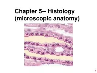
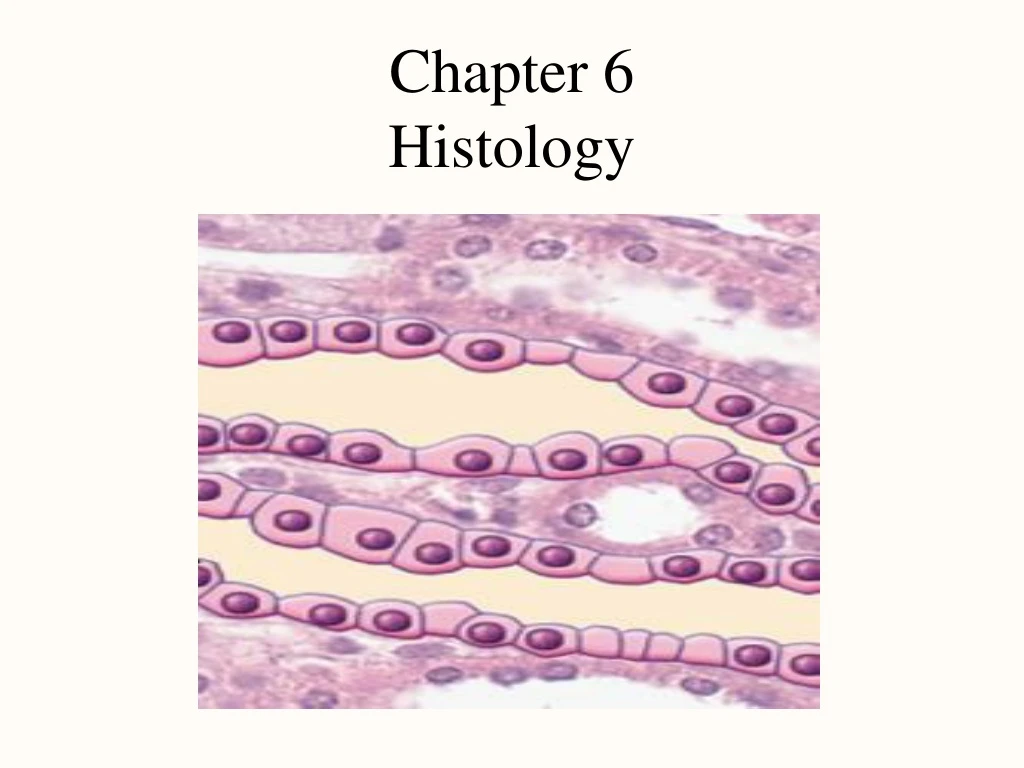
Chapter 6 Histology • Introduction -tissues a) multicullar structure – tissues contain cells of similar type. The cells work together to perform a specific function. b)cells and matrix – 1) matrix- extracellular material containing protein and mineral salts. 2) fluid surrounding cells is the interstitial fluid (Inter - between cells vs. Intra – inside a cell)
Chapter 6 Histology 2) Embryonic tissues- tissues from which all other tissues will arise. Starts with fertilized egg. Forms 3 layers (strata) called the primary germ layers. a) ectoderm (outer) -forms epidermis & nervous system b) endoderm (inner)-forms mucous membrane lining GI tract & respiratory system and digestive glands c) mesoderm (middle)-forms mesenchyme that gives rise to muscle, bone, blood and other connective tissues
Chapter 6 Histology 3) 4 basic tissue types (adult) – a) epithelial 1) one or more cell layers thick, free surface 2) simple or stratified 3) coverings –protection, secretion absorption
Chapter 6 Histology 3) 4 basic tissue types (adult) – b) connective 1) extensive extracellular matrix; collagen (flexible but strong), elastin (elastic qualities) 2) support, binding, protective 3) Connective tissue proper- Loose (adipose (fat)) tissue, Areolar (loosely binds epithelial cells to deeper tissue, and Reticular (supportive framework for lymphatic organs.
Epithelial Tissue There are two major categories of epithelial tissue: membranous and glandular. Membranous: The outer layer of skin, inner lining of cavities and lumina and coverings of visceral (internal-especially the heart, lungs, liver, pancreas or intestines) organs. Glandular: specialized tissue that form secretory portions of glands.
Membranous- Simple Epithelia Simple Epithelial Tissue: single layer thick and is located where diffusion, absorption, filtration and secretion are principle functions. Some cells contain cilia that move materials across cell surfaces. Other cells contain microvilli that increase surface area for absorption.
Membranous- Simple Epithelia Simple Squamous Epithelium : Flattened , irregularly shaped cells with an oval centrally located nucleus. Write down three places in the body where each type of epithelial cell can be found.
Membranous- Simple Epithelia Simple Columnar Epithelium : Tall, columnar cells with a single nucleus located near the basement membrane. Specialized unicellular glands called Goblet cells are scattered through this tissue. Goblet cells secrete a lubricative and protective mucus. Write locations.
Membranous- Simple Epithelia Simple Cuboidal Epithelium: Tightly fitted cube shaped cells found lining the lumina of small ducts and tubules that have excretory, secretory and absorptive functions. Write locations.
Membranous- Simple Epithelia Simple Ciliated Columnar Epithelium: Similar to columnar cells but with cilia along the free surface. Cilia do what? Where are they located?
Membranous- Simple Epithelia Pseudostratified Ciliated Columnar Epithelium: Ciliated columnar epithelium that appears to be layered- but each cell is anchored to the basement membrane. Write location.
Membranous- Stratified Epithelia Stratified Epithelia : Tissue consisting of two or more layers. This tissue is primarily protective and enhanced by rapid cell divisions. Stratified epithelia are classified according to the shape of the surface layer of cells. Why is this tissue not well suited for absorption and secretion?
Membranous- Stratified Epithelia Stratified Squamous Epithelium: Composed of a variable number of cell layers that are flattest at the surface. Mitosis occurs at only at the deepest layers. Keratinized: Contains keratin (strengthening protein) Nonkeratinized: No keratin, called mucosa Where are they located?
Membranous- Stratified Epithelia Stratified Cuboidal Epithelium: Consists of two or three layers of cuboidal cells where stratification provides a more robust lining. What location?
Membranous- Stratified Epithelia Transitional Epithelium: similar to nonkeratinized stratified squamous epithelium. Where is this located and what is the specific function?
Chapter 6 Histology Body Membranes: Thin layers of epithelial tissue that cover, support and protect visceral organs and line body cavities. Mucous Membrane: secrete thick mucus to lubricate and protect associated organs. Oral and nasal cavities, respiratory, reproductive, urinary and digestive system. Serous Membrane: secrete serous fluid to help protect. Pleurae (lungs), Pericardial (heart), Peritoneal (adominopelvic)
Glandular Epithelia Glandular Epithelia: Tiny invaginations (infoldings) of membranous epithelia that give rise to specialized secretory structures called exocrine glands. Unicellular: Single cells (Example ?) Multicellular: These can be simple or compound and are composed of both secretory cells and cells that form the walls of ducts.
Membranous- Stratified Epithelia Multicellular Exocrine Glands: Are organized the way they release their product. Merocrine- exocytosis through cell membrane Apocrine- secretion accumulates near surface of the cell and then is released with a portion of the cell. Holocrine- the entire secretory cell is discharged along with secretory product.
Chapter 6 Histology 3) Connective cont. Dense regular (tendons and ligaments), Dense irregular (dermis- forms protective coverings around organs, bones, nerves and most cartilages), Elastic (large arteries, lower respiratory tract, between arches of vertebrae)
Chapter 6 Histology Connective continued • Cartilage- consisting of chondrocytes, provides support along with elastic/flexible properties. Bone- comprised of spongy bone and compact or dense bone Blood- fluid connective tissue
Cartilage Hyaline cartilage: eases joint movement, holds airway open during respiration, growth zones of long bones in children.
Cartilage Elastic cartilage: Abundant elastic fibers that provide flexibility. Found in framework of outer ear, auditory canal and portions of larynx.
Cartilage Fibrocartilage: Abundant collagenous fibers that withstand compression. Found in the symphysis pubis, intervertebral discs, and knee joint.
Bone Bone: The most rigid connective tissue. Unlike cartilage, bone has a rich vascular supply and is the site of metabolic activity. Bones density is due mostly to inorganic calcium phosphate (calcium hydroxyapatite). Spongy (porous) (think Spongebob) or Compact (dense)
Blood Blood: vascular tissue or fluid connective tissue that helps to maintain homeostasis. Contains plasma, erythrocytes (red blood cells), leukocytes (white blood cells), and thrombocytes (platelets).
Question Review Why do some injuries to tendons and ligaments require more time to heal than a broken bone. Use your book and notes to formulate a reasonable answer- you may use smart phones at this time. (10 minutes)
Chapter 6 Histology 3) 4 basic tissue types (adult) – c) muscle 1) skeletal, cardiac, smooth 2) contractile 3)excitable – membrane potential – electrical charge
Skeletal Muscle Skeletal muscle: attaches to bone and allows voluntary movement. Muscle cells are multinucleated, striated, cylindrical fibers.
Cardiac Muscle Cardiac muscle: branched, striated fiber with a single nucleus and intercalated discs (gap junctions).
Smooth Muscle Smooth muscle: Elongated, spindle fibers with a single nucleus. Involuntary movement of internal organs.
Review Question Why are skeletal muscle cells multinucleated and why does cardiac muscle have intercalated disks? Use your book and notes to formulate a reasonable answer- you may use smart phones at this time. (10 minutes)
Chapter 6 Histology 3) 4 basic tissue types (adult) – d) nervous 1) Consits of neurons and neuroglia (or glial cells) 2) glia – provide protection for neurons neurons – nerve cells 3) Rapid signaling 4) Highly excitable – membrane potential
Chapter 6 Histology Tissue growth and repair a) embryonic tissue differentiates into adult tissue types. b) growth 1) hyperplasia – incr. cell # . 2) hypertrophy - incr. cell size. c) shrinkage /death 1) atrophy- decr. in cell size 2) necrosis – premature tissue death 3) infarction – sudden death to interrupted blood supply. a) cell rupture, stimulated inflammation. 4) apoptosis – programmed cell death a) cell shrinks, phagocytized.