Download
1 / 84
1.2k likes | 3.96k Views
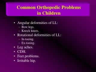
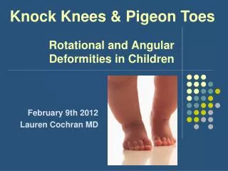
Knock Knees & Pigeon Toes. Rotational and Angular Deformities in Children. February 9th 2012 Lauren Cochran MD. To-Do. Search PREP for more questions? Email Myra to ensure no Mac-to-PC glitches? Summary slide / reasons to refer Clean up references slide. Objectives.

E N D
Knock Knees & Pigeon Toes Rotational and AngularDeformities in Children February 9th 2012 Lauren Cochran MD
To-Do • Search PREP for more questions? • Email Myra to ensure no Mac-to-PC glitches? • Summary slide / reasons to refer • Clean up references slide
Objectives • To review the common causes of intoeing & outtoeing in children • To describe the progression of normal physiologic alignment over time & how to evaluate a child with genu varum or genu valgum • To describe the physical examination techniques used in assessing rotational and angular deformities • To review indications for imaging and/or Orthopedics referral
4 Categoriesof Lower Extremity Problems • Rotational Deformities • Intoeing (“Pigeon Toes”) • Outtoeing • Angular Deformities • Genu Varum (“Bow legs”) • Genu Valgum (“Knock knees”) • Foot Deformities (e.g., clubfoot & pes planus) • Hip Disorders (e.g., DDH & SCFE)
Intoeing • Often raised as concern by parents • Sometimes associated with tripping/falling • Possible origin: • Foot = metatarsus adductus • Between knee & ankle = internal tibial torsion • Between hip & knee = medial femoral torsion
Metatarsus Adductus (MTA) • “Packaging Defect” (1st born children at higher risk) • Tarsal & phalangeal bones angled toward midline • + convex lateral curvature of foot • + medial instep crease • Evaluation includes: • Degree of adduction (heel bisector) • Degree of flexibility
Metatarsus Adductus • Heel bisector (quantifies degree of adduction): • Normal: between 2nd/3rd toes • Mild MTA: 3rd toe • Moderate MTA: 3rd/4th toe • Severe MTA: 4th/5th toe
Metatarsus Adductus • Degree of Flexibility determines need for treatment! • Actively correctable: tickle foot on lateral border no treatment required • Passively correctable: gentle lateral pressure to 1st metatarsal head “stretching” 10sec per foot x5 with each diaper change REFER if not corrected by 4-6mo • Rigid: REFER for possible casting/bracing
Metatarsus Adductus & DDH? • Previously reported association between MTA & DDH now disputed • Still warrants careful hip exam
Clubfoot • aka talipes equinovarus • Constellation of metatarsus adductus PLUS: • Equinus positioning = inability to dorsiflex foot (shortening of gastroc/soleus & tendon) • Hindfoot varus (supination) • Inversion of the forefoot • Warrants early referral to Orthopedist • Famous athletes with clubfoot: Troy Aikman, Kristi Yamaguchi, Mia Hamm
Internal Tibial Torsion • Origin of intoeing between knee & ankle • Most common intoeing etiology for children less than 3yrs • “Packaging defect” present from birth but often noticed only when child begins to stand/walk • Almost never requires treatment: • Most cases gradually resolve by age 2-3yrs • REFER if persists beyond age 6
Medial Femoral Torsion • Origin of intoeing between hip & knee • aka “femoral anteversion” but to a pathologic degree (> 60-65) • Most common intoeing etiology for children older than 3yrs • Associated with “W sitting position” (unclear effect on progression/outcome) • “Egg beater” or “windmill” running pattern • Almost always corrects gradually by age 10
Intoeing: H&P • HPI: onset, progression, pain, disability, previous evaluation/treatment • Development: particularly gross motor milestones including walking • General screening exam: • Growth parameters • Evaluation for DDH (Ortolani/Barlow, Galeazzi, skin fold asymmetry) • Basic neurological exam (UE+LE reflexes, ankle clonus, heel/toe walking)
Intoeing: “Rotational Profile” • Foot progression angle • Heel bisector • Thigh foot angle • Hip rotation
Foot Progression Angle • Normal = -5 to +20 • Mild intoeing: -5 to -10 • Moderate intoeing: -10 to -15 • Severe intoeing: > -15
Thigh Foot Angle • Patient prone with knees flexed to 90 degrees + natural foot position • Line down center of thigh to heel bisector • Normal = 0 to +10-15 • Negative = internal tibial torsion
Hip Rotation • Child is prone with knees flexed to 90 • Assess both sides simultaneously • Normal (varies by age): ~-45 to +45
Hip Rotation • If asymmetric get XR (AP pelvis AP) to evaluate for DDH or other hip problem • Increased internal rotation > 60-65= femoral anteversion • > 80 = moderate • > 90 = severe
Benefits of Intoeing? • Some evidence that persistent intoeing is actually beneficial for some sports that require quick directional shifts: • Tennis • Basketball • Soccer • Etc.
Outtoeing • Much less common than intoeing • Origin: • Foot: Calcaneovalgus deformity • Between knee & ankle: External tibial torsion • Between hip & knee: Femoral retroversion • Hip: External rotation contracture • Most cases resolve within first 1-2yrs of ambulation
Calcaneovalgus Foot • Hyperdorsiflexion of foot • Abduction of forefoot • Forefoot often rests on anterior surface of leg • REFER for casting if foot cannot be plantar-flexed below neutral
External Tibial Torsion • Thigh foot angle > +20 • Possible compensation for femoral anteversion • REFER for severe cases (> 40) • REFER if persistent > age 6yrs