Download
1 / 80
800 likes | 808 Views
Explore the diverse functions and crucial role of proteins in living organisms, including their amino acid composition, chiral nature, and essential amino acids.

E N D
CHAPTER 11:Proteins: Structure and Function OUTLINE 11.1 Amino Acids 11.2 Chirality and Amino Acids 11.3 Peptides 11.4 Protein Architecture 11.5 Enzymes
CHAPTER 11:Proteins: Structure and Function BIOMOLECULES Biomolecules are large, complex organic molecules. They are present in all living things. They include: Proteins Carbohydrates Lipids Nucleic acids (DNA and RNA) The first of these is presented below, the remainder in subsequent sets of slides. All obey general principles of chemical reactions discussed previously.
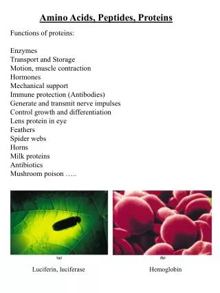
CHAPTER 11:Proteins: Structure and Function PROTEINS Proteins have some of the most diverse functions of all biological molecules, ranging from the hemoglobin that transports oxygen to tissues, to collagen and elastin that provide structure to ligaments, tendons, and blood vessels, to the enzymes that catalyze all biochemical reactions. They are composed of amino acids linked together in chains, folded into complex structures with specific biological functions.
CHAPTER 11:Proteins: Structure and Function PROTEIN FUNCTION IS DEPENDENT ON STRUCTURE! The change in a single component of the protein hemoglobin results in altered folding patterns for the protein, which affects cellular structure resulting in a serious disease, sickle cell anemia.
CHAPTER 11:Proteins: Structure and Function DID YOU KNOW? 1. What is the chemical basis for sickle cell anemia? 2. How does molecular polarity play a role in hemoglobin function?
CHAPTER 11:Proteins: Structure and Function DID YOU ANSWER? Hemoglobin, like all globular proteins, has surface polar amino acids and internal nonpolar amino acids. In sickle cell anemia, one of its surface amino acids, glutamic acid, is genetically exchanged for the nonpolar amino acid, valine. This simple shift results in the incorrect conformation, or folding, of the protein, causing the disease.
CHAPTER 11:Proteins: Structure and Function 11.1 AMINO ACIDS
CHAPTER 11:Proteins: Structure and Function RECALL Chemical properties of organic functional groups Effect of pH on acidic and basic groups
CHAPTER 11:Proteins: Structure and Function PARTS OF AMINO ACIDS An amino acids consists of an amine a carboxylic acid a hydrogen atom one of about 20 side chains (R groups or residues) All bonded to a central atom, the a-carbon The identity of an amino acid is determined by its side chain.
CHAPTER 11:Proteins: Structure and Function AMINO ACID STRUCTURE AND pH The amino group is basic; the carboxylic acid group is acidic. At neutral pH, each is ionized. Compounds such as amino acids containing both a negative and a positive charge are called zwiterions. As the pH of an amino acid solution changes, its charges change.
CHAPTER 11:Proteins: Structure and Function AMINO ACID SIDE CHAINS The 20 amino acids are differentiated by their side chains. The side chains may be either polar or nonpolar. Nonpolar side chains are generally alkanes or have aromatic ring structures. Polar side chains may be divided into acidic, basic, or neutral side chains. At physiological pH, the 3 basic side chains have positive charge the 2 acidic side chains have negative charge
CHAPTER 11:Proteins: Structure and Function EXAMPLES OF AMINO ACIDS Aspartic acid has an acidic side chain, lysine has a basic side chain, and cysteine has a neutral side chain. Each amino acid may be designated by a 3-letter abbreviation, as shown.
CHAPTER 11:Proteins: Structure and Function NONPOLAR AMINO ACIDS *Essential Amino Acid
CHAPTER 11:Proteins: Structure and Function POLAR AMINO ACIDS
CHAPTER 11:Proteins: Structure and Function ESSENTIAL AMINO ACIDS Essential amino acids are amino acids required in our diets. About half may be synthesized by our bodies, the rest must be consumed in the foods we eat. Essential amino acids include the following:
CHAPTER 11:Proteins: Structure and Function PRACTICE PROBLEM For each of the following amino acids check the boxes that apply.
CHAPTER 11:Proteins: Structure and Function 11.2 CHIRALITY AND AMINO ACIDS
CHAPTER 11:Proteins: Structure and Function AMINO ACID CHIRALITY Chirality is a property associated with many molecules, particularly those with biomedical applications such as most amino acids. Objects, such as a glove or certain molecules, are chiral if their mirror images are non-superposable. Your right and left hands have chirality because they are mirror images, and yet are not the same as each other. The term “chiral” means “handed.” Objects, such as a sock or mitten, are achiral because their mirror images are superposable. Chiral molecules have at least one tetrahedral atom with four different atoms or groups attached.
CHAPTER 11:Proteins: Structure and Function ENANTIOMERS Enantiomers are pairs of chiral objects, one being the mirror image of the other. Amino acids may consist of enantiomers. But only one of the pair is found in nature. It is customary to refer to each member of the enantiomeric pair with the prefixes D- or L-. Only the L-form of amino acids is common. Other common classes of materials are in the D-form. Other naming rules may use R- and S, (+) and (-), or d-, and l-.
CHAPTER 11:Proteins: Structure and Function EXAMPLE: A PAIR OF ENANTIOMERS The common form of alanine is the L-form. Each has the same set of groups bonded to the a-carbon, but in a different 3-dimensional relationship Note the mirror-image relationship between them:
CHAPTER 11:Proteins: Structure and Function FISCHER PROJECTIONS A simplified way to indicate the structure of enantiomers is to use Fischer projections. These indicate chiral atoms as crosshairs (+) with the main carbon chain written vertically. For L-amino acids, the alpha carbon is shown at the crosshair with the carboxylate to the top, the amino group to the left and the side chain down. D-amino acids would be the mirror image of this.
CHAPTER 11:Proteins: Structure and Function ALANINE AS A FISCHER PROJECTION
CHAPTER 11:Proteins: Structure and Function PROPERTIES OF ENANTIONMERS When enantionmers are in an achiral environment, their physical and chemical properties are identical. In a chiral environment, such as within a cell, their properties may be quite different. Because proteins are composed of L-amino acids, they are chiral. For example two common drugs, Darvon and Novrad are enantiomers, but one is an analgesic and the other is a cough suppressant.
CHAPTER 11:Proteins: Structure and Function A PAIR OF ENANTIONMERS These pharmaceuticals are mirror images of each other, yet they show very different effects in our bodies.
CHAPTER 11:Proteins: Structure and Function RACEMIC MIXTURES A racemic mixture is a mixture containing both enantiomers, the L- form and the D-form. Since one of the pair usually does not have biological activity, the racemic mixture is likely to have reduced potency. These are usually designated by indicating both forms, such as D/L. In many cases, the enantiomer without biological activity displays adverse effects if consumed. Most pharmaceuticals contain only one of the two enantiomers.
CHAPTER 11:Proteins: Structure and Function PRACTICE PROBLEMS 1. Indicate whether the following statements are TRUE or FALSE. A chiral molecule: a. is superposable on its mirror image b. has an enantiomer c. may exhibit different chemical properties from its enantiomer in the body d. typically exhibits different chemical properties from its enantiomer in an achiral environment 2. Ibuprofen, the active ingredient in Motrin and other OTC analgesics, is a chiral drug sold as a racemic mixture. What does this mean?
CHAPTER 11:Proteins: Structure and Function 11.3 PEPTIDES
CHAPTER 11:Proteins: Structure and Function PEPTIDE BONDS Peptides are molecules formed by joining two or more amino acids. Peptides may be small oligopeptides (2 amino acids to about a dozen) larger polypeptides (up to about 50 amino acids) very large molecules called proteins (>50 amino acids) To form these, amino acids are joined by peptide bonds, essentially an amide group. Peptide bonds form between the carboxylate group of one amino acid and the ammonium on another.
CHAPTER 11:Proteins: Structure and Function PEPTIDE FORMATION The formation of a peptide bond is an example of an acyl group transfer reaction. The reverse is a hydrolysis. Note the involvement of a water molecule in these reactions.
CHAPTER 11:Proteins: Structure and Function OLIGOPEPTIDES Two amino acids in a peptide are a dipeptide. A third joined to these constitute a tripeptide. Structures of any length may be formed. One end of the chain will always have an ammonium ion at neutral pH, and is called the N-terminus; the opposite end is a carboxylate ion and is called the C-terminus.
CHAPTER 11:Proteins: Structure and Function A SPECIFIC TRIPEPTIDE The tripeptide shown consists of the amino acids alanine, glycine, and valine. Alanine is the N-terminal amino acid and valine is the C-terminal amino acid. Peptides are conventionally written with the N-terminus to the left.
CHAPTER 11:Proteins: Structure and Function COMMON OLIGONUCLEOTIDES Aspartame, commercially the artificial sweetener Nutrasweet™, is the dipeptide asp-phe. Oxytocin, a hormone involved in contractions during labor and in lactation, consists of the 9 amino acids: cys - tyr - ile - gln - asn - cys - pro - leu – gly The endorphin, met-enkephalin, mentioned in chapter 7, has 5 amino acids. This helps reduce pain following injury.
CHAPTER 11:Proteins: Structure and Function PRACTICE PROBLEM Are the dipeptides gly-phe and phe-gly different compounds? Are they structural isomers or stereoisomers?
CHAPTER 11:Proteins: Structure and Function 11.4 PROTEIN ARCHITECTURE
CHAPTER 11:Proteins: Structure and Function PROTEIN STRUCTURE Proteins are composed of the 20 L-amino acids. A protein may have from about 50 to many thousands of amino acids, joined linearly by way of peptide bonds. The information for determining the sequence of amino acids resides in the DNA of most cells. A gene is the region of DNA responsible for the coding of a protein. There are thousands of different proteins in the body, each has a specific function dependent on its structure.
CHAPTER 11:Proteins: Structure and Function THREE-DIMENSIONAL SHAPE OF PROTEINS Proteins are not strictly linear structures. The chain of amino acids folds into a three-dimensional structure termed its native conformation. Electrostatic interactions between atoms within the protein and between protein atoms and external atoms, such as solvent, determine folding patterns. Amino acid side chains contribute interacting groups to this folding.
CHAPTER 11:Proteins: Structure and Function FOUR LEVELS OF PROTEIN STRUCTURE Primary structure Sequence of amino acids from N-terminus to C-terminus Secondary structure Localized regular folding stabilized by hydrogen bonds Tertiary structure Complex irregular folding of entire protein Quaternary structure Association of two or more subunits
CHAPTER 11:Proteins: Structure and Function PRIMARY STRUCTURE OF A PROTEIN The sequence of amino acids determines all other aspects of a protein’s structure and function. If this sequence is altered, the protein may not function properly. Genetic disease is usually a disruption of the primary structure, often by replacing a single amino acid. In sickle cell anemia, a single amino acid change from glutamic acid to valine results in improper function of hemoglobin. An amino acid is located in sequence by the number of its position from the N-terminus.
CHAPTER 11:Proteins: Structure and Function SECONDARY STRUCTURE Secondary protein structure refers to the regular folding patterns in localized regions of a protein. Most interactions stabilizing secondary structure occur between carbonyl oxygens and amide hydrogens by way of hydrogen bonds:
CHAPTER 11:Proteins: Structure and Function PATTERNS IN SECONDARY STRUCTURE There are many examples of extended patterns of secondary structure. The two most common types of secondary structure are a-helix b-pleated sheet Typical proteins may contain some of each of these or be mostly one or the other; some proteins contain neither.
CHAPTER 11:Proteins: Structure and Function THE a-HELIX An a-helix is a coiled segment of a polypeptide held in place by hydrogen bonds between carbonyl and amide groups along the protein backbone.
CHAPTER 11:Proteins: Structure and Function RIBBON DRAWING OF a-HELIX The diagrams show a portion of the oxygen-binding protein, myoglobin. The ribbons on the left indicate the seven regions of a-helix. The two ribbons in red are expanded on the right to show hydrogen bonds.
CHAPTER 11:Proteins: Structure and Function THE β-PLEATED SHEET b-pleated sheets occur when two or more polypeptide chains stack in a pattern similar to that of a pleated skirt. These are stabilized by hydrogen bonding, and the chains may be either parallel to each other (each chain in the same orientation N-terminal to C-terminal) or antiparallel (chains in opposite orientation). The strength of silk is due to the presence of b-pleated sheets in the protein fibroin.
CHAPTER 11:Proteins: Structure and Function THE β-PLEATED SHEET Ribbon drawings of b-pleated sheets usually indicate these structures with broad flat arrows, pointing in the direction of the C-terminal amino acid.
CHAPTER 11:Proteins: Structure and Function PRACTICE PROBLEMS Orient the peptide structures below to show how they would form hydrogen bonds, and add dashes to show the corresponding hydrogen bonds. Label the partial positive and negative charges in the atoms involved in hydrogen bonding. Why are there partial charges?
CHAPTER 11:Proteins: Structure and Function TERTIARY STRUCTURE The tertiary structure of proteins describes the entire folding pattern of the protein and consists of complex and irregular folding, not patterned as found in secondary structure. Tertiary structure is determined by interactions that include distant amino acid residues as well as the surrounding environment. For some proteins, tertiary structure also includes prostheticgroups—non-peptide organic molecules or metal ions that are strongly bound to the protein.
CHAPTER 11:Proteins: Structure and Function EXAMPLES OF TERTIARY STRUCTURE
CHAPTER 11:Proteins: Structure and Function FOUR LEVELS OF PROTEIN STRUCTURE Primary structure Sequence of amino acids from N-terminus to C-terminus Secondary structure Localized regular folding stabilized by hydrogen bonds Tertiary structure Complex irregular folding of entire protein Quaternary structure Association of two or more subunits
CHAPTER 11:Proteins: Structure and Function WHAT CAUSES A PROTEIN TO FOLD? The fundamental driving force behind protein folding is energy. A folded protein and its environment are lower in potential energy than the unfolded protein. Proteins fold spontaneously into their native conformation under physiological conditions. Protein folding is not random, but is defined by the sum of all the electrostatic interactions present in the protein and its surroundings.
CHAPTER 11:Proteins: Structure and Function TYPES OF ELECTROSTATIC INTERACTIONS IN PROTEINS There are 4 main types of electrostatic interactions resulting in the tertiary structure of a protein: Disulfide bridges (covalent bond, hydrophobic) Salt bridges (ionic bond, hydrophilic) Hydrogen bonding (hydrophilic) Dispersion forces (hydrophobic)