Download
1 / 28
310 likes | 947 Views
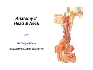
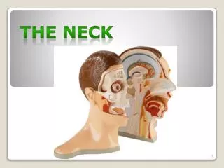
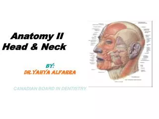
Anatomy II Head & Neck BY: DR.Yahya Alfarra CANADIAN BOARD IN DENTISTRY. Parotid gland :: Size : it’s the largest salivary gland & it’s compose of serous acini. Shape : it’s wedge shaped , Site & extension : it lies between the ramus of Md. & SCM It extends :

E N D
Anatomy II Head & Neck BY:DR.Yahya AlfarraCANADIAN BOARD IN DENTISTRY
Parotid gland :: Size : it’s the largest salivary gland & it’s compose of serous acini. Shape : it’s wedge shaped , Site & extension : it lies between the ramus of Md. & SCM It extends : Upward: to zygomatic arch Downward: to angle of Md. Anteriorly: to cover part of masseter ms. Posteriorly: to overlap SCM Parotid region
Length : 5 cm long Beginning : at the ant. Border of the gland End: by opening into the oral cavity opposite the upper 2nd molar tooth Parotid duct(stenson’s duct)
Opens in the oral cavity opposite to the second upper molar tooth.
Blood supply of P.G • Arterial supply : small branches from ECA inside the gland • Venous drainage: into retromandibular V. • Lymphatic drainage : into deep & superficial parotid lymph node • Nerve supply of parotid :Sensory : great auricualr N. auriculotemporal N.
Temporal region • Temporal fossa • Infratemporal fossa • MuSclE OF mastications
Boundaries : Sup. Temporal line ……… above Zygonatic arch…………….below Frontal process of zygomatic bone……………………….ant. Contents(attachments): 2 superficial Ms of mastications Muscles : a) temporalis Ms. b) masseter Ms Nerves : a ) deep temporal N. b)auriculotemporal N. Vesseles: superficial temporal A. Temporal fossa
Boundaries: Anteriorly: post. Surface of maxilla Posteriorly : styloid & mastoid process Medially: lateral pterygoid plate Laterally : coronoid process & ramus of Md. Contents: Muscles : 2 deep Ms of Mastications a) medial pterygoid Ms. b ) lateral pterygoid m. Nerves: a ) Md. N. b ) chorda tympani c ) Mx. N. Vesseles : a ) Mx. A. b ) pterygoid plexus of viens Infratemporal fossa
Submandibualr region • Definition: it’s the regoin between Md. & hyoid bone. • It contains : • Muscles: digastric, mylohyoid , hyoglossus, geniohyoid , genioglossus & styloglossus • Salivary gland : submandibualr & sublingual • Nerves : lingual , glossopharyngeal & hyoglossal • Blood vesseles: facial A. & V. , Lingual A. & V. • Parasympathetic ganglion : submandibualr • Lymph nodes : submandibualr group
Site & extension: It lies below Md. It extends to : Mylohyoid line : a bove Digastric muscle : below Mental foramen : anterior Angle of Md. : posterior Size: about ½ the size of P.G. Shape: wedge shape Structure: it consists of 2 parts,a large superficial part & a small deep part Submandibular gland
It’s 5 cm long It’s duct open into the floor of mouth close to frenulum of tongue. Submandibular duct
Blood supply of gland: • The arterial supply:the gland is supplied by branches of facial & lingual A. • The viens drain into facial & lingual V. • Nerve supply : • sympathetic :from plexus arround lingual A. • Parasympathetic:from facial N.
Site: beneath tongue Size: it’s the smallest of the 3 S.G. Shape: almond-shaped Relation : Ant. : the gland of the opposite side Post. : the deep part of submandibualr gland Med. : genioglossus Ms. , lingual N. & submand. Duct Laterally: sublingual fossa of Md. Sup. : mucous membrane of mouth(which elevated to form sublingual fold) Inf. : mylohyoid Ms. Sublingual gland
Sublingual duct : It opens directly in the floor of mouth on sublingual fold • Blood supply :the gland is supplied by branch of facial & lingual A. The viens drain into facial & lingual V. • Nerve supply : • sympathetic :from plexus arround lingual A. • Parasympathetic:from facial N.
Nerves OF submandibualr regoin lingual , glossopharyngeal & hyoglossal N.B. : lingual N. : branch of post. Division of Md. N. in the infratemporal fossa • Blood vesseles submandibualr regoin : facial A. & V. , Lingual A. & V. N.B. Facial A. : Branch of ECA Facial V. : It units ant. Division of retromandibualr V. TO drain into IJV Lingual A. : Branch of ECA Lingual V. : drain into IJV