Download

1 / 30
340 likes | 800 Views
Male Reproductive System. Metallic 0 Mind Bilal Marwa Sarah Al- Morit. TESTIS. Covered by Tunica Vaginalis , Covered by:simple squamous mesothelium , surround the anterolateral aspect of the testis.

E N D
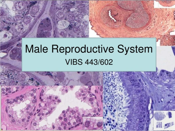
Male Reproductive System Metallic 0 Mind BilalMarwa Sarah Al-Morit
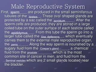
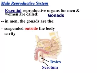
TESTIS • Covered by Tunica Vaginalis, • Covered by:simplesquamousmesothelium, • surround the anterolateral aspect of the testis. Testis is surrounded by a capsule of dense, irregular collagenous connective tissue known as Tunica Albugenia (white color) Deep to tunica albugenia is a highly vascularized connective tissue tunica vasculosa
Mediastinum testis sends septa to subdivide the testis into 250 pyramid- shaped compartment known as lobuli testis Tunica vasculosa thickens in the posterior aspect to form mediastinum testis. # seminiferous start blend opened in the tubular structure (rete testis)
SEMINIFEROUS TUBULES • epithelium: • Several layers thick. • Two types of cells: • Sertoli cells (somatic) • Spermatogenic cells • tunica propria (C.T.): • Collagen type I • Fibroblasts • In animals other than human myoid cells are present, they are similar to smooth muscle cells. • Well developed basal lamina Company Logo
SERTOLI CELLS Functions? physical and nutritional suport for germ cells Phagocytosis: excess cytoplasm (spermiogenesis) establishment of blood testis barrier synthesis of androgen binding protein (ABP), antimüllaerian hormone, inhibin and testicular transferrin. secretion of fructose-rich medium Where? • Lie on basal lamina Company Logo
SERTOLI CELLS STRUCTURE A- pale basophilic B-basally located oval nucelus with large central nuceolus. C- inclustions: crystalloids of Charcot-Böttcher. D- SER, limited RER, golgi apparatus, numerous mitochondria and vesicles and abundant cytoskeletal elements E- abundant lysosomes F- phagocytose excess cytoplasm in spermiogenesis Company Logo
Sertoli cells form occludent junctions between them which divide the lumen of seminiferous tubule into apical tooccludent junctions Sertoli cells cannot divide after puberty 1- Adluminal compartment 2- Basal compartment Basal to occluden junctions Blood-testis barrier: formed by junctions between sertoli cells. Function:protect devloping gametes from the immune system
SPERMATOGENIC CELLS Spermatogonia: • Located in basal compartment. • Diploid cells Company Logo
SPERMATOGENIC CELLS • Priamryspermatocytes, secondary spermatocytes, spermatids and spermatozoa occupy the adluminal compartment. • Primary speramatocytes: • Largest cells. • Large vesicular nuclei. • Secondary spermatocytes: • Short-lived. • Do not replicate . • Enter the second mitotic division. Company Logo
Spermiogenesis • Spermatids: small round haploid cells • Spermiogenesis: spermatids spermatozoa Company Logo
Spermatozoon HEAD: • Surrounded by plasma lemma. • Contains nucleus • Acrosome surrounds the nucleus MIDPIECE: • Contains the mitochondrialsheath • encircles the axoneme and outer dense fibers. Company Logo
Spermatozoon TAIL • Longest segment. • Contains axoneme surrounded by 7 outer dense fibers END PIECE • Composed of axoneme surrounded by plasmalemma Company Logo
Contents of a lobule:2- connective tissue • Loose, vascularized connective tissue. • Contains: fibroblasts, mast cells and other things in loose connective tissue • (read epithelium and connective tissue lecture) • Also contains interstitial cells (leydig cells) Company Logo
Interstitial cells (Leydig cells) • Polyhedral in shape • Single, vesicular nucleus. • May be binucleated. • Mitochondria with tubular cristae. • SER and golgi apparatus. • Some RER. • Numerous lipid droplets. • No secretory vesicles. • Crystals of Reinke. Company Logo
GENITAL DUCTS Company Logo
Intratestsicular Ducts • Tubulirecti: • Lined by sertoli cells in the first half. • The second half is lined by simple cuboidal epithelium • Rete testis: • Lined by simple cuboidal epithelium Company Logo
DuctuliEfferentes: • Conduct the sperm from rete testis to epididymis. • Lining:nonciliatedcuboidal cells alternating with the regions of cilitaed columnar cells • Has basal lamina and connective tissue and thin layer of smooth muscle Company Logo
Epididimis • The lumen is lined by psuedostratified epithelium with 2 cells • Basal Cell • short pyramidal cells with round nuclei (heterochromatin). • They are stem cells • Principal cells: • Tall • RER,SER, golgi apparatus multivesicular bodies • have stereocilia which are nonmotilemicrovilli Company Logo
Epididimis • Has a basal lamina which separates the epithelium from loose connective tissue • Has a layer of smooth muscle cells Company Logo
Vas (ductus) deferans • Epithelium: • Stereociliatedpseudostratified columnar epithellium • Basal lamina • Loose fibroelastic connective tissue • has numerous folds • Smooth muscle has 3 layers : • Inner and outer longitudinal and middle circular • loose fibroelastic connective tissue • Ampulla: Has a highly folded, thickended epithelium Company Logo
Seminal Vesicle • Mucusa is highly convoluted • No stereocilia • Epithelium: • Pseudostratified columnar epithelium with 2 types of cells: 1- basal cells 2- columnar cells • Subepithelial C.T. is fibroelastic Company Logo
Seminal vesicle Smooth Muscle layers: 1- inner circular 2- outer longitudinal Smooth muscle cells surrounded by fibroelastic connective tissue Company Logo
Prostate • Capsule: • dense,irregularcollagenous C.T. interspersed with smooth muscle cells. • Prostate composed of compound tubolaveolar glands arranged into: • Mucosal • Submucosal • Main Company Logo
Prostate • Mucosal glands • Closest to the urethra • Shortest of the glands • Submucousalglands: • Peripheral to mucousal glands. • Larger than mucosal glands. • Main Glands: • Largest and most numerous of the glands • Compose the bulk of the prostate Lumina of the glands contains prostatic concretions (corpora amylacea) composed of calcified glycoprotein. Company Logo
C.T. stroma of the gland: • Derived from the capsule and has the same structure • So it is enriched by smooth muscle fibers (+ C.T.) • Lining of the gland components: • Simple to psuedostratified columnar epithelium • Secretion: serous Company Logo
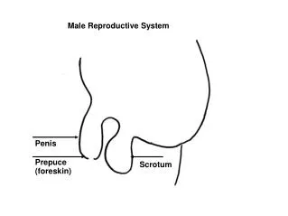
PENIS • Composed of 3 columns of erectile tissue • each column is enclosed by its own capsule: Tunica Albuginea: Dense, fibrous C.T. • The columns are: • 2 corpora carnevosa • Located dorsally • Tunica albuginea not continuous • 1 corpus spongiosum • Located ventrally • Houses penile urethra • Ends as glans penis Company Logo
PENIS • The three columns are surrounded by a common sheath • Loose C.T. • No hypodermis • Erectile tissue: • Endothelially lined spaces • Seperated by trabeculae of connective tissue and smooth muscle cells and elastic fibers Company Logo
PENIS Company Logo