STOMACH
470 likes | 808 Views
Discover the intricate details of the stomach anatomy, from the cardiac sphincter to the gastric glands, and learn how these components work harmoniously to aid in digestion and nutrient absorption. Explore the complex structure of the stomach's layers and cell types to appreciate its vital role in maintaining overall health.

STOMACH
E N D
Presentation Transcript
STOMACH Dr IramTassaduq
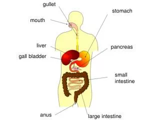
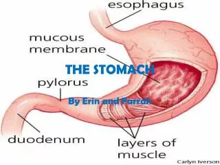
STOMACH • The stomach functions both as a reservoir and as a digestive organ. It empties its contents in small portions (suitable for continued digestion) into the small intestine
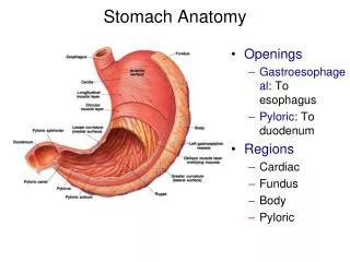
Anatomically, the stomach is divided into • cardiac part, • fundus, • body • pyloric part (pyloric antrum and pyloric canal)
CARDIAC SPHINCTER • The cardiac sphincter is a specialized valve found between the esophagus and the stomach. It prevents backflow of food and digestive enzymes.
FUNDUS • The is the frontal region of the stomach. It begins digestion of proteins and mixes together stomach contents.
BODY • The body is the central region of the stomach. It also digests proteins and blends materials found in stomach.
PYLORUS and PYLORIC SPHINCTER • The pylorus is the back region of the stomach. It contracts to empty materials from the stomach into the small intestine • The pyloric sphincter is a specialized valve that prevents materials and digestive enzymes from escaping into the small intestine before digestion is completed in the stomach.
RUGAE LINING OF THE STOMACH • Rugae are found on the interior layer of the stomach and aid in breaking down food when the stomach contracts.
Gastric areas (mammillated areas). Mucosa is divided by furrows into small irregular elevations. These are the gastric areas. Gastric pits (foveolae). surface of each gastric area to be studded with minute depressions, the gastric pits.
Mucosa. • Epithelium Simple columnar • Lamina Propria contains glands which differ in each histological region of the stomach. • Muscularis Mucosae. It measures from 0.3-1.5 mm in thickness, being thinnest in the cardia and thickest in the body and fundus.
SUBMUCOSA • This layer separates the muscularis from the mucosa . • It consists of coarse collagenous fibers and many elastic fibers, plus blood vessels, lymph vessels, nerves, and the plexus of Meissner. Glands are absent.
MUSCULARISEXTERNA • an outer longitudinal, • a middle circular, • and an inner oblique. • The oblique layer is best developed in the cardia and body. The circular layer is thickest in the pylorus where it forms the pyloric sphincter, which helps control the evacuation of food. The longitudinal layer is continuous with the longitudinal muscle layer of the esophagus and duodenum. The myenteric plexus lies in the connective tissue lamina, which separates the circular from the longitudinal muscle fibers.
SEROSA • The outer layer consists of loose connective tissue covered on its superficial aspect by mesothelium. Small blood vessels, lymphatics, and nerves lie in the connective tissue.
SURFACE EPITHELIUM • surface epithelium (simple, tall columnar). • It contains mucus-producing cells, surface mucous cells, • The mucus is alkaline and adheres to the epithelium. which protects the mucosa from the acidic contents of the stomach.
GASTRIC GLANDS • cellular composition and function of the gastric glands are specialized in the different parts of the stomach
GASTRIC GLANDS - Secrete mucus to protect epithelial cells from enzymes & acid - Secrete HCl (for protein digestion) & intrinsic factor (for B12 absorption) - Secrete pepsinogen which gets converted to “pepsin” when mixed with HCl; for protein digestion - Secrete gastrin to regulate stomach emtying Entero-
CARDIAC GLANDS • Cardiac glands are heavily branched tubular glands which contain mainly mucus-producing cells. • Shallow Gastric Pits • Short glands
PRINCIPAL or FUNDIC GLANDS • Each glandular tubule consists of three parts: • deep body, • intermediary neck • upper isthmus.
PYLORIC GLANDS • Branched, coiled tubular glands. • Endocrine cells, in particular gastrin-producing cells, are more frequent than in principal glands. A few parietal cells may be present but chief cells are usually absent.
In the pylorus, the pits extend downward to about one-half the thickness of the mucosa; • in the cardia, and fundus, the pits occupy only one-fourth the thickness of the mucosa
MUCUS NECK CELLS • are found between the parietal cells in the neck of the gland.They are difficult to distinguish from chief cells in plain H&E stained section
Chief cells (or zymogenic cells) • contain Zymogens granules • Abundant RER give the cell Basophilic stain • most numerous of the four types. They occur primarily in the body of the glands. • produce pepsinogen, which is a precursor of the proteolytic enzyme pepsin.
Parietal cells (or oxyntic cells) • Large acidophilic cells. • secrete the hydrochloric acid of the gastric juice. Aside from activating the pepsinogen • Parietal cell also secrete intrinsic factor, which is necessary for the resorption of vitamin B12
On electron microscopy reveal Intracellular canalicular system and Tubulovesicular System
ENTEROENDOCRINECELLS • Scattered between the epithelial cells of the gastric mucosa and their basement membrane. • Demonstrated by Electron microscope or by Immunological technique.On light microscopy they appear clear. • Secretion is released in the lamina propria and distributed by blood vessels.
Endocrine cells • The best characterized endocrine cells in the gastric mucosa are gastrin-producing cells (G cells) and somatostatin-producing cells (D cells). G cells are most frequent in the middle third of the glands. They stimulate the secretion of acid and pepsinogen. G cell function is stimulated by nervous input, the distension of the stomach. • D cells are found mainly in glands of the pyloric antrum. They inhibit G cells and thereby acid production. D cell function is stimulated by acid in the lumen of the stomach and duodenum.
Cell renewal • The surface epithelium is renewed approximately every third day. • The source of the new cells is the isthmus, i.e. the upper part of the neck, of the gastric glands, where cells divide and then migrate towards the surface epithelium and differentiate into mature epithelial cells