Download
1 / 1
10 likes | 98 Views
Diffusion Image Analysis. Martha Shenton Ph.D. ‡ , Marek Kubicki M.D. Ph.D. ‡ , Sylvain Bouix Ph.D. ‡ ,

E N D
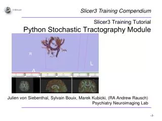
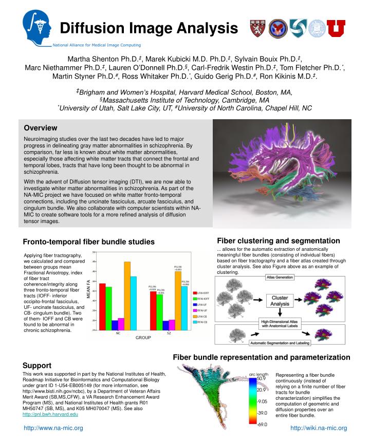
Diffusion Image Analysis Martha Shenton Ph.D.‡, Marek Kubicki M.D. Ph.D.‡, Sylvain Bouix Ph.D.‡, Marc Niethammer Ph.D.‡, Lauren O’Donnell Ph.D.§, Carl-Fredrik Westin Ph.D.‡, Tom Fletcher Ph.D.*, Martin Styner Ph.D.#, Ross Whitaker Ph.D.*, Guido Gerig Ph.D.#, Ron Kikinis M.D.‡. ‡Brigham and Women’s Hospital, Harvard Medical School, Boston, MA, §Massachusetts Institute of Technology, Cambridge, MA *University of Utah, Salt Lake City, UT, #University of North Carolina, Chapel Hill, NC Overview Neuroimaging studies over the last two decades have led to major progress in delineating gray matter abnormalities in schizophrenia. By comparison, far less is known about white matter abnormalities, especially those affecting white matter tracts that connect the frontal and temporal lobes, tracts that have long been thought to be abnormal in schizophrenia. With the advent of Diffusion tensor imaging (DTI), we are now able to investigate whiter matter abnormalities in schizophrenia. As part of the NA-MIC project we have focused on white matter fronto-temporal connections, including the uncinate fasciculus, arcuate fasciculus, and cingulum bundle. We also collaborate with computer scientists within NA-MIC to create software tools for a more refined analysis of diffusion tensor images. Fiber clustering and segmentation ... allows for the automatic extraction of anatomically meaningful fiber bundles (consisting of individual fibers) based on fiber tractography and a fiber atlas created through cluster analysis. See also Figure above as an example of clustering. Fronto-temporal fiber bundle studies Applying fiber tractography, we calculated and compared between groups mean Fractional Anisotropy, index of fiber tract coherence/integrity along three fronto-temporal fiber tracts (IOFF- inferior occipito-frontal fasciculus, UF- uncinate fasciculus, and CB- cingulum bundle). Two of them- IOFF and CB were found to be abnormal in chronic schizophrenia. Fiber bundle representation and parameterization Support This work was supported in part by the National Institutes of Health, Roadmap Initiative for Bioinformatics and Computational Biology under grant ID 1-U54-EB005149 (for more information, see http://www.bisti.nih.gov/ncbc), by a Department of Veteran Affairs Merit Award (SB,MS,CFW), a VA Research Enhancement Award Program (MS), and National Institutes of Health grants R01 MH50747 (SB, MS), and K05 MH070047 (MS). See also http://pnl.bwh.harvard.edu Representing a fiber bundle continuously (instead of relying on a finite number of fiber tracts for bundle characterization) simplifies the computation of geometric and diffusion properties over an entire fiber bundle. http://www.na-mic.org http://wiki.na-mic.org