Download
1 / 34
360 likes | 424 Views
Understand the significance of familial pheochromocytoma in genetic disorders and its clinical implications. Learn about symptoms, diagnosis factors, and hereditary syndromes associated with this rare tumor.

E N D
Familial pheochromocytoma BitaMirzaei MD Endocrinology FellowResearch Institute for Endocrine sciencesShahidBeheshti University of Medical Sciences Mordad 94
Silent pheochrmocytoma? Familial pheochromocytoma? Isolated familial pheochromocytoma? Genetic evaluation?
Among patients with adrenal incidentalomas, approximately 5.1% to 6.5%, even up to 23%, proved to have pheochromocytomas. And 10% of adrenal pheochromocytomas have presented as adrenal incidentalomas, even with clinically silent. Pheochromocytomas are rare tumors found in less than 1% of the populations with hypertension. Although the majority of patients are symptomatic, 10-30% of pheochromocytomas are clinically silentCrout and Sjoerdsm found that pheochromocytomas 50gm. or larger are often asymptomatic because secreted catecholamines are metabolized within the tumor. In contrast, tumors small than 50 gm. have slow turnover rates and release free cateholamines into the circulation, exhibiting persistent symptoms and signs
For pheochromocytomas, the mean diameter of incidental tumors was also significantly larger than that of symptomatic tumors.
Of all the adrenal pheochromocytoma, 20% to 30% of them are asymptomatic; they are called clinically silent pheochromocytoma. • Generally, the neoplasm shows a larger tumour size at the time of diagnosis because of the delay to the first visit • Different factors contribute to the diagnosis: • (1) extensive necrosis cystic region at the centre of the mass may significantly decrease the number of cells producing catecholamine (2) interstitial tissue without bioactivity may be the main ingredient of the neoplasm; considerable blood sinus around the cystic area is also demonstrated • (3) most of the catecholamines and metabolic product may be stored in the capsular mass, which infuses intravenously into the blood circulation when isolating the mass; as such, a hypertension crisis is possible.
PHEOCHROMOCYTOMA IN GENETIC DISORDERS • Most catecholamine-secreting tumors are sporadic. However, approximately 30 percent of patients have the disease as part of a familial disorder; in these patients, the catecholamine secreting tumors are more likely to be bilateral adrenal pheochromocytomas or paragangliomas. • Hereditary catecholamine-secreting tumors typically present at a younger age than sporadic neoplasms.
There are several familial disorders associated with pheochromocytoma, all of which have autosomal dominant inheritance: • von Hippel-Lindau (VHL) syndrome, MEN2 and, less commonly, neurofibromatosis type 1 • approximate frequency of pheochromocytoma in these disorders is 10 to 20 percent in VHL syndrome, 50 percent in MEN2, and 0.1 to 5.7 percent with neurofibromatosis type 1
Pheochromocytomas also occur with paraganglioma type 1 , type 2, type 3 type 4 and type 5 ; which are caused by mutations in thePheochromocytomas also occur with paraganglioma type 1 , type 2, type 3 type 4 and type 5 ; which are caused by mutations in the SDHD SDHAF2 SDHC SDHB and SDHA genes, respectively.
VHL syndrome — The VHL phenotype includes pheochromocytoma (frequently bilateral), paraganglioma (mediastinal, abdominal, pelvic), hemangioblastoma (involving the cerebellum, spinal cord, or brain stem), retinal angioma, clear cell renal cell carcinoma, pancreatic neuroendocrine tumors, endolymphatic sac tumors of the middle ear, serous cystadenomas of the pancreas, and papillary cystadenomas of the epididymis and broad ligament. • The VHL tumor suppressor gene, located on chromosome 3p25-26, encodes a protein that regulates hypoxia-induced proteins. More than 300 germline VHL mutations have been identified that lead to loss of function of the VHL protein.
Patients with VHL syndrome may be divided into two groups: type I and type II • Patients with type I syndrome do not develop pheochromocytoma, whereas patients with type II syndrome are at high risk for developing pheochromocytoma. • In addition, type II VHL syndrome are subdivided into type IIA (low risk for renal cell carcinoma), type IIB (high risk for renal cell carcinoma), and type IIC (pheochromocytomas only) • . Genotype-phenotype correlations have been documented for this disorder and specific mutations are associated with particular patterns of tumor formation
MEN 2A is characterized by medullary thyroid cancer (MTC) in all patients, pheochromocytoma in 50 percent, primary hyperparathyroidism in 20 percent, and cutaneous lichen amyloidosis in 5 percent • MEN type 2B represents approximately 5 percent of all MEN2 cases and the phenotype is characterized by MTC in all patients, pheochromocytoma in 50 percent, mucocutaneousneuromas (typically involving the tongue, lips, and eyelids) in most patients, skeletal deformities (eg, kyphoscoliosis or lordosis), joint laxity, myelinated corneal nerves, and intestinal ganglioneuromas
Phenotype of MEN2 versus VHL syndrome — The clinical and biochemical characteristics of pheochromocytomas in MEN2 versus the VHL syndrome were investigated in a study of 19 and 30 patients with these disorders, respectively; the following findings were noted: • MEN2 patients were more symptomatic with a higher incidence of hypertension (primarily paroxysmal). • MEN2 patients all had elevated serum concentrations of metanephrine (the epinephrine metabolite), while all VHL patients had elevated serum normetanephrine concentrations (the norepinephrine metabolite. • Compared to MEN2 tumors, VHL tumors had lower total tissue contents of catecholamines and expression of tyrosine hydroxylase (TH), the rate-limiting enzyme in catecholamine synthesis. They also had much lower expression of phenylethanolamine N-methyltransferase (PNMT, the enzyme that converts norepinephrine to epinephrine) and tissue stores of epinephrine. • Thus, the difference in clinical phenotype (MEN2 patients are more symptomatic) can be explained by the differences in biochemical phenotype (MEN2 patients, due to the higher PNMT and TH expression, have an adrenergic phenotype with higher rates of catecholamine biosynthesis.
MTC is generally the first manifestation of MEN 2A. • Probands with MTC typically present with a neck mass or neck pain, usually before age 35 years. Up to 70% of such individuals already have cervical lymph node metastases. All individuals with an MTC-predisposing mutation who have not undergone prophylactic thyroidectomy demonstrate biochemical evidence of MTC by age 35 years.
Pheochromocytomas usually present after MTC or concomitantly; they are the first symptom in 13–27% of individuals with pheochromocytomas and MEN 2A. • Pheochromocytomas in persons with MEN 2A and MEN 2B are diagnosed at an earlier age, have subtler symptoms, and are more likely to be bilateral than sporadic tumors
Pheochromocytoma was the only clinical manifestation of their syndrome in 38% of carriers of von Hippel‑Lindaudisease and 24% of carriers of MEN2.Pheochromocytoma was the only clinical manifestation of their syndrome in 38% of carriers of von Hippel‑Lindaudisease and 24% of carriers of MEN2.
Neurofibromatosis type 1 • Approximately 2 percent of patients with NF1 develop catecholamine-secreting tumors. In these patients, the catecholamine-secreting tumor is usually a solitary benign adrenal pheochromocytoma, occasionally bilateral adrenal pheochromocytoma, and rarely a peri adrenal abdominal paraganglioma. • Genetic testing for NF1 is available but is not routinely performed, as the diagnosis is made based upon clinical phenotype.
Other mutations in 2010, loss of function mutations in the FP/TMEM127 gene were identified in patients with familial and sporadic pheochromocytoma, but not paraganglioma. • In a study of 990 individuals with pheochromocytoma or paraganglioma, germline mutations in FP/TMEM127 were identified in 20 individuals with adrenal tumors, five of whom had a family history of pheochromocytoma • Among 547 patients who presented with sporadic pheochromocytoma (unilateral adrenal tumor with negative family history), 11 had FP/TMEM127 mutations. • In 2011, loss of function mutations in the MAX gene were identified in patients with familial pheochromocytoma . • In an initial study of three individuals with familial pheochromocytomaMAX germline mutations were found.. • In an extension of this study, MAX mutations were found in 5 of 59 patients (8.5 percent) with suspected familial pheochromocytoma (based on age of onset <30 years, bilateral pheochromocytoma, or positive family history)
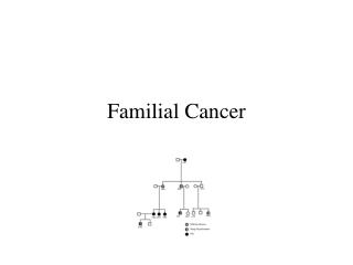
VHL syndrome in a Turkish family of 49 members from three generations was evaluated, ten patients had malignant and benign tumors compatible with VHL syndrome 20 % of the family members had pheochromocytoma, 8 % had renal cell carcinoma, while 6 % had pancreatic neuroendocrine tumor, 2 % retinal angioma, and 2 % had spinal hemangioblastoma. In nine of our patients we observed pheochromocytoma. Mean age of patients was 34 years (range 24–55). • In five patients, mass was located in bilateral adrenal glands.
This report supports the few previous accounts of isolated familial pheochromocytoma occurring in individuals shown to carry mutations in the VHL gene (Crossey et al 1995, Walther and Linehan 1996, Ritter et al 1996). In contrast to the other reported mutations, the S68W mutation appears to present with variable penetrance of isolated pheochromocytoma
Suggested approach — Given the considerable cost of genetic testing, using a stepwise approach based on each patient's clinical scenario is prudent • The First International Symposium on Pheochromocytoma concluded that it is not cost effective to test for every disease-causing gene in every case, and suggested that the secretion profile, patient age, focality of primary tumor, and prior history be included in a genetic testing algorithm
Recommendation 3.2 • We recommend the use of a clinical feature-driven diagnostic algorithm to establish the priorities for specific genetic testing in PPGL patients with suspected germline mutations. (1QQQE)
GENETIC SCREENING — Genetic testing should be considered if a patient has one or more of the following: • Paraganglioma • Bilateral adrenal pheochromocytoma • Unilateral adrenal pheochromocytoma and a family history of pheochromocytoma/paraganglioma • Unilateral adrenal pheochromocytoma onset at a young age (eg, <45 years), or • Other clinical findings suggestive of one of the previously discussed syndromic disorders • An asymptomatic person at risk for disease on the basis of family history of pheochromocytoma/paraganglioma should have genetic testing only if an affected family member has a known mutation.