Sense Organs: The ye & the Ear
550 likes | 1.03k Views
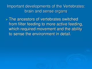
Sense Organs: The ye & the Ear. THE EAR. Combining Forms for the ear: ot/o, aur/o , auricul/o Two functions of the ear: Hearing Equilibrium (balance) Three separate regions of the ear: Outer ear Middle ear Inner ear. OUTER EAR.

Sense Organs: The ye & the Ear
E N D
Presentation Transcript
THE EAR • Combining Forms for the ear: ot/o, aur/o, auricul/o • Two functions of the ear: Hearing Equilibrium (balance) • Three separate regions of the ear: Outer ear Middle ear Inner ear
OUTER EAR • Auricle (pinna) –projecting flap where sound waves enter • Auditory canal leads from pinna to middle ear • Produces cerumen (ear wax) which lubricates and protects the ear Auricle Auditory canal
MIDDLE EAR Sound waves travel through auditory canal and strike membrane between outer & middle ear. This membrane is called the tympanic membrane or eardrum. The tympanic membrane vibrates with sound waves and moves 3 small bones in the middle ear. These small bones are called ossicles. Tympanic Membrane
MIDDLE EAR The three small bones also have individual names: • Malleus – the TM moves this bone first • Incus – vibration moves this bone next • Stapes (staped/o) – vibrates last and touches the next membrane called the oval window Incus Malleus Tympanic Membrane Stapes
MIDDLE EAR Oval window separates the middle ear from the inner ear. Eustachian Tube (salping/o) leads from the middle ear to pharynx • Is normally closed, except when swallowing. • Aids in prevention of damage to eardrum and shock to middle and inner ear when air pressure is greater in the middle ear than in atmospheric air. Incus Malleus Tympanic Membrane Stapes Eustachian Tube Oval Window
INNER EAR Also called the labyrinth because of it’s circular, maze-like structure. Cochlea (cochle/o) leads from the oval window; small, snail shaped; has two parts: 1. Auditory Liquids a. Perilymph b. Endolymph c. Vibrations travel through these liquids 2. Organ of Corti – sensitive auditory receptor; are tiny hair like calls that receive vibrations from auditory liquids and relay sound waves to auditory nerve fibers. Cochlea
INNER EAR Auditory Nerve Fibers – receive sound vibrations from the auditory liquids and end in the auditory center of the cerebral cortex of the brain where impulses are heard and interpreted. Vestibule – connects the cochlea to 3 semicircular canals for balance. Semicircular canals (Organ for Equilibrium) – contain endolymph & hair cells that fluctuate in response to movement of the head; nerve fibers send message to brain & brain sends message to muscles to maintain balance. “-saccule” & “–utricle” are membranous sacs Auditory Nerve Fibers Vestibule Semicircular Canals Cochlea
SEQUENCE OF EVENTS IN STIMULATION OF SENSE ORGAN Figure 17-1. Pattern of events in the stimulation of a sense organ.
PATHWAY OF SOUND VIBRATIONS Figure 17-21. Pathway of sound vibrations from the outer ear to the brain (cerebral cortex).
TYPES OF DEAFNESS • Conduction – caused by impairment of the middle ear ossicles and membranes that transmit sound waves into cochlea. • Nerve – caused by impairment of the cochlea or auditory nerve
AURAL TEMPERATURE Figure 17-25. Ear thermometer using a tympanic membrane thermometer.
COMBINING FORMS & ABBREVIATIONS acous/o = HEARING acoustic = audi/o = SENSE OF HEARING audiometer = audit/o = HEARING auditory = -cusis = HEARING presbycusis = -otia = EAR CONDITION macrotia = LARGE EARS microtia = SMALL EARS ABBREVIATIONS AD = RIGHT EAR AS = LEFT EAR AU = BOTH EARS ENT = EAR, NOSE, & THROAT EENT =EYES, EARS, NOSE, & THROAT
PATHOLOGICAL CONDTIONS 1. Otitis Media – inflammation of middle ear, caused by infection (staphylococcus or streptococcus bacteria) 2. Tinnitus – tinkling sound in the ear; can be ringing, buzzing, whistling, etc…. 3. Vertigo – sensation of irregular or whirling motion of oneself or external objects; equilibrium and balance are affected.
TYMPANIC MEMBRANE • Healthy tympanic membrane. • Tympanic membrane with cholesteatoma. • Tympanic membrane with acute otitis media. • Myringotomy with tympanostomy tube.
PATHOLOGICAL CONDTIONS 4. Meniere’s Disease – disorder of labyrinth marked by elevated endolymph pressure. Symptoms include tinnitus, vertigo, and loss of hearing. Cause is unknown. Bedrest, sedation and drugs for nausea and vertigo are commonly given. 5. Otosclerosis – bone growth around oval window and stapes leading to fixation (stiffening) causing improper conduction of vibration. Corrected with stapedectomy and replacement by prosthesis.
TREATMENT FOR A FORM OF CONDUCTION HEARING LOSS • Stapedectomy. Using microsurgical technique and a laser, the stapes bone is removed from the middle ear. • A prosthetic device (wire, Teflon, or metal) is placed into the incus and attached to a hole in the oval window.
DEFINE THESE PATHOLOGICAL CONDITIONS Otomycosis - ot/o = __ myc/ = __ osis = Myringotomy - myring/o = ___ tomy = Myringitis - myring/o = __ itis = __ Otopyorrhea - ot/o = __ py/o = __ rrhea = Ossiculoplasty - ossicul/o = ___ plasty =
EAR PROCEDURES Audio / gram – Audio / meter – Audio / metry – Oto / scopy –
AUDIOMETER Figure 17-24. Pure-tone audiometer.
OTOSCOPIC EXAM Figure 17-26. Otoscopic examination. The auricle is pulled up and back. The hand holding the otoscope is braced against the face for stabilization.
STRUCTURES OF THE EYE Cor/o and Pupil/o = pupil Pupil - the dark center of the eye Conjunctiv/o – conjunctiva Conjunctiva – clear membrane that lines inner surface of eyelids & over whites of eyes. Corne/o and kerat/o = cornea Cornea – a fibrous, transparent tissue that extends over the pupil of the eye and iris. Helps with refraction of light. Conjunctiva Cornea Pupil
STRUCTURES OF THE EYE Scler/o – sclera Sclera – white part of the eye. Avascular – no blood vessels. Choroid – dark brown membrane inside sclera. Choroid Sclera
STRUCTURES OF THE EYE Ir/o – iris Iris – colored portion of the eye. • Is also a muscle that surrounds pupil of the eye. • If light is bright, the iris gets bigger (contracts) & pupil gets smaller (constricts). • If light is dim, the iris relaxes & pupil dilates (gets larger). Mi/o – smaller, less Miosis: constriction of the pupil Mydr/o – wider, enlarge Mydriasis: enlargement of pupil Iris Ciliary Body
STRUCTURES OF THE EYE Cycl/o – ciliary body Ciliary body – a muscle located next to the lens and that two functions: 1. Thickens & thins the lens for refraction (bending of light rays) 2. Secretes aqueous humor (AH) Aqueous Humor – fluid that maintains shape of anterior portion of eye & provides nourishment to structures in the same area. Ciliary Body
STRUCTURES OF THE EYE Phak/o and phac/o – lens of the eye Lens – flat for distant vision and rounded for close vision. Also helps with refraction of light. Refractive power of lens is ACCOMODATION Lens
STRUCTURES OF THE EYE Anterior Chamber of Eye: • Contains aqueous humor • Secreted by ciliary body • Maintains shape of anterior eye • Constantly produced - leaves eye thru canal that carries it to bloodstream Anterior Chamber
STRUCTURES OF THE EYE Vitreous Chamber of Eye: • Contains vitreous humor • Maintains shape of posterior eye • Refracts light rays • Is NOT constantly being produced • Loss of VH may mean loss of eye Vitreous Chamber
STRUCTURES OF THE EYE Retin/o - retina Retina – thin, nerve layer that contains rods and cones. • Rods are for reduced lighting and peripheral vision. • Cones are for color and central vision. Retina
STRUCTURES OF THE EYE • When light energy hits rods & cones (in retina) causes chemical changes, that initiate nerve impulse to travel to brain via the optic nerve • Optic disc is area where optic nerve meets retina (it has no light receptors – so called blind spot) Optic Disc Optic Nerve Fovea Centralis Macula
STRUCTURES OF THE EYE • Macula is small area to the side of the optic disc • Macula contains the Fovea Centralis: location of sharpest vision within eye (composed largely of cones) Optic Disc Optic Nerve Fovea Centralis Macula
RETINA Figure 17-3. The posterior, inner part (fundus) of the eye, showing the retina as seen through an ophthalmoscope.
BINOCULAR VISION • Optic Nerve Fiber – carry light stimulus through nerve fibers to the brain. • As fibers enter brain, travel more medially & eventually cross. • Optic Chiasm – area where optic nerve fibers cross • Nerve fibers from right 1/2 of each retina form an optic tract & synapse in thalamus. • Fibers end in the right visual field of the cerebral cortex. • Same thing happens with the left half of each retina • Images fuse, giving 3 dimensional image: called Binocular Vision
PATHWAY OF LIGHT Figure 17-5. Pathway of light rays from the cornea of the eye to the cerebral cortex of the brain.
AREAS OF BRAIN INVOLVED IN VISION • Thalamus – nerve fibers from right half of each retina form an optic tract and synapse in the thalamus. Fibers will end in the right visual field of the cerebral cortex. Same thing happens with nerve fibers on the left half of each retina. • Cerebral Cortex – surface of cerebrum where nerve cells lie in sheets. Receive visual stimulus from thalamus from both sides of the eye. • Visual area of cerebral cortex is in occipital lobe of the brain.
VISIONAL DISTURBANCES • Accommodation – normal adjustment of the eye for seeing objects at various distances – eye has more problems with this as ages • Astigmatism – defective curvature of the cornea or lens of the eye. • Presby/opia – impaired vision of the cornea or lens of the eye, associated with aging. • Hyper/opia – farsightedness (can’t see close objects), rays of light focus behind retina • My/opia – nearsightedness (can’t see far away objects), rays of light focus in front of the retina
ERRORS OF REFRACTION • Astigmatism – defective curvature of the cornea or lens of the eye. • Presby/opia – impaired vision of the cornea or lens of the eye, associated with aging. • Hyper/opia – farsightedness (can’t see close objects), rays of light focus behind retina • My/opia – nearsightedness (can’t see far away objects), rays of light focus in front of the retina • Astigmatism and its correction. • Hyperopia and its correction. • Myopia and its correction. Dashed lines in B and C indicate the contour and size of the normal eye.
COMBINING FORMS Ambly/o = DULL, DIM Amblyopia = Dipl/o = DOUBLE Diplopia = Nyct/o = NIGHT Nyctalopia = Phot/o = LIGHT Photophobia = -opsia = VISION Hemianopsia = Blephar/o = EYELID Blepharoptosis = Dacry/o = TEAR DUCTS Lacrim/o = TEARS Lacrimal ducts =
LACRIMAL TEAR DUCTS Lacrimal (tear) gland and ducts.
ABBREVIATIONS OD – RIGHT EYE OS – LEFT EYE OU – BOTH EYES VF – VISUAL FIELD AH – AQUEOUS HUMOR VH – VITREOUS HUMOR IOP – INTRAOCULAR PRESSURE
DISORDERS OF THE EYE Figure 17-6. (A) Acute bacterial conjunctivitis. Notice the discharge of pus characteristic of this highly contagious infection of the conjunctiva. (B) Anisocoria.
DISORDERS OF THE EYE Cataract. The lens appears cloudy.
DISORDERS OF THE EYE Figure 17-10. Chalazion.
DISORDERS OF THE EYE Figure 17-11. Glaucoma and circulation of aqueous humor. Circulation is impaired in glaucoma, so that aqueous fluid builds up in the anterior chamber.
DISORDERS OF THE EYE Figure 17-12. (A) Picture as seen with normal vision. (B) The same picture as it would appear to someone with macular degeneration.
PROCEDURES A normal fluorescein angiogram.
PROCEDURES Figure 17-14. Ophthalmoscopy. In addition to examining the cornea, lens, and vitreous humor for opacities (cloudiness), the examiner can see the blood vessels at the back of the eye (fundus) and note degenerative changes in the retina.
PROCEDURES Figure 17-15. Slit lamp examination measuring intraocular pressure by tonometry.
PROCEDURES (A) The Snellen chart assesses visual acuity. (B) Visual fields are examined by comparing the patient's field of vision with that of the examiner's (assuming the examiner's is normal).