Download
1 / 63
630 likes | 645 Views
Comprehensive information on identification, diagnosis, and management of strokes and TIAs for medical students. Includes types, symptoms, risk factors, and treatment options. Vital knowledge for future practice.

E N D
Strokes and TIAs Ben Ryan Final Year Medical Student
Contents • Background Info • Definitions and Overview • Types of Strokes • Recognising Stroke • Stroke Mimics • Stroke • Diagnosing • Acute Management • Secondary Prevention • Sub-arachnoid haemorrhage • Central Venous Thrombosis • TIAs • DVLA • History Taking
Why it’s Important • Stroke/TIA history is a completely reasonable OSCE station • Explanation of Secondary Prevention of stroke has came up in the past • Important to know for future practice
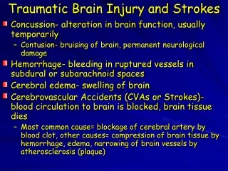
Stroke Summary • Rapidly developing clinical signs of focal (sometimes global) disturbance of cerebral function lasting more than 24 hours, or leading to death, with no apparent cause other than that of vascular origin • Caused by infarction or haemorrhage from one of the cerebral arteries • Clinical features are determined by location affected • Will likely need CT to determine which • If infarction (not haemorrhagic) – thrombolysis if within 4.5 hours, or 300mg Aspirin OD for 2 weeks • Some haemorrhagic strokes require neurosurgical treatment • 70% of strokes occur in those aged >70, but can occur at any age • Scores to be aware of: ROSIER (recognising stroke) and CHA2DS2-VASC (annual risk of stroke in those with AF and no anticoagulation) • FAST is for public use, not doctors
TIA Summary • Transient Ischaemic Attack • Stroke clinical features that resolve within 24 hours • Usually resolves within a couple of minutes, or a few hours at most • Anybody with continuing neurological signs when first assessed should be assumed to have a stroke • Should be assessed for future risk of stroke (ABCD2) • All should get aspirin 300mg daily started immediately and secondary prevention • If high risk: specialist assessment within 24 hours • If low risk: specialist assessment within 1 week
What’s the diagnosis? • 60 year old male presenting with left arm and leg weakness, left homonymous hemianopia and dysphasia, which started 4 hours ago and is still present
What’s the diagnosis? • 60 year old male presenting with arm and leg weakness with slurred speech which stopped after 15 minutes
What’s the diagnosis? • 60 year old male presenting with left homonymous hemianopia which has became gradually worse over the past month
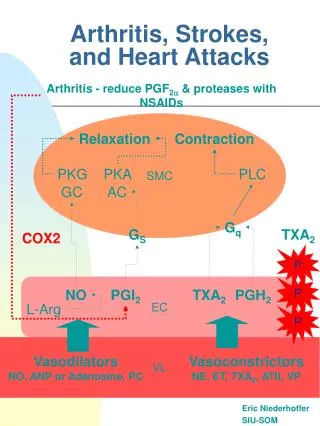
Aetiology • Cerebral Infarction/Ischaemic (88%) • Restricted or interrupted blood supply to an area of the brain • Thrombosis, secondary to atherosclerosis • Or cerebral embolism from AF, valve disease/replacement, post-MI, or DVT embolism with patent foramen ovale (paradoxical embolism) • Or an episode of hypoperfusion • Cerebral haemorrhage (12%) • Bleeding into an area of the brain • Intra-cerebral (9%) sounds is the typical haemorrhagic stroke we think of • Intra-parenchymal or intra-ventricular • Sub-arachnoid haemorrhage (3%) appears to officially be a type of haemorrhagic stroke • Distinguishing between the two is extremely important for management
Types of Strokes (Bamford/Oxford Classification) • Based on initial symptoms • Total Anterior Circulation Stroke • Partial Anterior Circulation Stroke • Lacunar Stroke • Posterior Circulation Stroke
Total Anterior Circulation Stroke (15%) • Stroke caused by middle/anterior cerebral arteries • All 3 of the following are required: • Unilateral weakness (and/or sensory deficit) of face, arm and leg (contralateral to stroke) • Homonymous hemianopia (contralateral to stroke) • Higher cerebral dysfunction (dysphasia, for example) • Problem with Anterior Cerebral Artery affects legs more than arms • Problem with Middle Cerebral Artery affects arms more than legs (arms are in the middle of the body)
Partial Anterior Circulation Stroke (25%) • Stroke from anterior circulation (anterior/middle cerebral arteries) • Two of the following: • Unilateral weakness (and/or sensory deficit) of face, arm and leg (contralateral to stroke) • Homonymous hemianopia (contralateral to stroke) • Higher cerebral dysfunction (dysphasia, for example)
Lacunar Strokes (25%) • Results from impairment of blood supply to the brain’s deep structures • Putamen, thalamus, caudate, pons, internal capsule • No evidence of higher cerebral dysfunction • One of: • Unilateral weakness (and/or sensory deficit) of face/arm/leg • Pure sensory symptoms on one side of the body • Ataxic hemiparesis (clumsiness/weakness on ipsilateral side)
Posterior Circulation Strokes (25%) • Involves vertebrobasilar arteries – supplies the occipital lobe, midbrain, brainstem and cerebellum • Presents with one of the following: • Ipsilateral cranial nerve palsy with contralateral motor and/or sensory deficit • Bilateral motor and/or sensory deficit • Disorder of conjugate eye movement • Cerebellar dysfunction • Isolated homonymous visual field defect • Basically, lots of different presentations. Most important to remember that it will affect cerebellum and brainstem.
Other patterns of stroke • Lateral medullary syndrome/Wallenburg’s syndrome (posterior inferior cerebellar artery) • Ipsilateral: ataxia, nystagmus, dysphagia, facial numbness, cranial nerve palsy • Contralateral: limb sensory loss • Weber’s syndrome (midbrain infarct) • Ipsilateral oculomotor nerve palsy and contralateral hemiplegia
Recognising • Sudden onset of neurological symptoms • FAST is for public use, not for doctor’s • Just covered the different stroke symptoms/syndromes • Think about: • Limb weakness/sensory disturbance • Facial weakness • Visual field defects • Difficulties with speech/swallowing • Ataxia, nystagmus • Cranial nerve palsies • ROSIER (Recognition of Stroke in the Emergency Room)
ROSIER • Loss of consciousness or syncope = - 1 point • Seizure activity = - 1 point • New acute onset of:Asymmetrical facial weakness = + 1 pointAsymmetrical arm weakness = + 1 pointAsymmetrical leg weakness = + 1 pointSpeech disturbance = + 1 pointVisual field defect = + 1 point • If score more than 0, acute stroke likely
Issues with ROSIER • Doesn’t assess for many features of posterior circulation strokes • Still use clinical judgement
Stroke Mimics and Stroke Chameleons • Diagnosing stroke is not always straight-forward • Stroke mimics are conditions which are not strokes, but may appear to be • Stroke chameleons are strokes that can be mistaken for other conditions • Stroke is common – high probability that sudden onset neurological deficit is a stroke
Stroke Mimics • Account for 20-25% of suspected stroke presentations: • Hypoglycaemia – always do a blood glucose • Hemiplegic migraine • Todd’s paresis • Sepsis • Brain Tumour • Metabolic • Neuropathies • Vestibular disorders • Extra/sub-dural haemorrhages • Drugs and alcohol
Todd’s paresis • Focal weakness in a part of a body after a seizure • Weakness may be similar to stroke • Usually subsides within 48 hours • Patients often have old strokes (not new) on imaging – not to be confused with an acute stroke • MRI with diffusion weighted imaging can help differentiate between old and new strokes
Hypoglycaemia • Can (rarely) present with focal neurological features • Are they on insulin, sulfonylureas, or recent high alcohol intake? • Important to always do blood sugars! • Airways, breathing, circulation, don’t ever forget glucose • ABCDEFG
Sepsis • Can mimic stroke • Can also co-exist with stroke, such as secondary to aspiration pneumonia • Septic screen will help
Migraine and other headache disorders • Headache is a common feature of acute ischaemic stroke (27%) • Some headache disorders can mimic stroke • Hemiplegic migraines is the most notable one • Symptoms will be reversible • Family history will help (autosomal dominant, high penetrance) • Typically young, female and have fewer attacks as they age • Difficult to diagnose on first presentation • CT/MRI usually normal
Brain Tumours • Will usually have progressive history over months of focal neurological features • May present with acute neurological features • Imaging should find it
Stroke Chameleons • Actual strokes that mimic other diseases, sometimes due to their tempo of onset (gradually progressive or stuttering) or symptoms don’t match typical arterial territory • Main benefit in diagnosing these is to start secondary prevention treatment (will likely be too late to give thrombolysis etc.) • Vertigo • Monoplegia • Delirium • Cauda Equina Syndrome
Vertigo • Acute onset of vertigo, dizziness and vomiting • Most people would either think Meniere’s or BPPV, depending on the history • However, strokes can present with vertigo (posterior circulation strokes)
Monoplegia • One limb weakness • Might be easy to think a problem with spinal cord • Could be an anterior circulation stroke
Delirium • Delirium • Acute onset and fluctuating course of disturbed mind: altered consciousness levels, disorganised thinking, inattention • Could be a stroke, affecting frontal lobe
Cauda Equina Syndrome • Typical features of cauda equina syndrome (acute onset back pain, lower limb weakness and paraesthesia, urinary retention/incontinence, faecal incontinence) • One of your differentials for this syndrome is spinal infarction • Should be seen when you scan the back
Summary • Have a low-threshold for considering stroke in any neurological findings • Be aware of your stroke mimics, and remember to do a blood glucose!
After recognising stroke - summary • A to E assessment as always • Establish whether haemorrhagic or ischaemic – brain imaging • If ischaemic, can you haemolyse? If not, high-dose aspirin • Most haemorrhagic strokes don’t need surgery, but some do • Strokes can interfere with blood pressure: • Need to be careful with blood pressure, it is often not necessary to lower it • Might need to lower it if giving thrombolysis, or in intracerebral haemorrhage • Swallowing assessment: trained nurses and doctors can do an admission swallowing screen • If this indicates problems, NBM until SALT assessment • Optimise nutrition in the mean time • All patients should be admitted to a stroke unit
Investigations • FBC • ESR • U&E • Blood glucose • Coagulation Screen • ECG • CXR • Cardiac monitor • Brain Imaging
Brain Imaging • Need to establish if haemorrhagic or ischaemic • All will need a scan – some will need immediately, some as soon as possible • Indications for immediate scan: • Candidate for thrombolysis • On anticoagulant treatment • Known bleeding tendency • Decreased GCS • Unexplained progressive or fluctuating symptoms • Papilloedema, neck stiffness or fever • Severe headache at onset of symptoms • Our trust guidance says CT is the brain imaging to do (I’m under the impression that diffusion weight MRI is better, but probably less accessible)
Management of Acute Ischaemic Stroke • Alteplase (thrombolysis) is recommended if it can be started within 4.5 hours of onset of symptoms • Check for contra-indications • Given over 1 hour. 10% as a bolus, other 90% as an IV infusion. • Risk of bleeding - Immediate CT if any worsening neuro function, and 24 hours after thrombolysis • If can not give thrombolysis, aspirin 300mg stat, and once daily for 14 days • This is also done 24 hours after thrombolysis • If can not take aspirin, use alternative anti-platelet such as clopidogrel or dipyridamole • Rarely, decompressive hemicraniectomy can be performed (if severe stroke and signs of an infarct in the MCA territory)
Other considerations • Ischaemic stroke and in AF? • Treat with aspirin 300mg for 2 weeks as per before considering anticoagulation treatment • For long-term secondary prevention of stroke • Clopidogrel 75mg daily is first line • Aspirin + dipyridamole is second line (or just one of them, if they are intolerant to the other) • Control of other risk factors • High-intensity statin – atorvastatin 20-80mg • Consider anti-hypertensives • Smoking, diabetes • Carotid imaging • More than 70% stenosis – surgery indicated
A little bit on Atrial Fibrillation • Atrial Fibrillation greatly increases the risk of strokes (can fire off embolisms) • For those with AF, can use CHA2DS2-VASC to calculate their annual risk of having a stroke • This is also used to help determine whether they should be on long-term anticoagulation, such as warfarin or the DOACs
Intracerebral Haemorrhage Management • A to E approach • Coagulation screen – reverse any increased anti-coagulation factors • Lower systolic BP to <140 • Some require surgical removal of haemorrhage • Deteriorating neurologically • Brainstem compression • Ventricular obstruction/hydrocephalus • Apparently has worse prognosis • Less evidence and guidance out there on this topic
Sub-Arachnoid Haemorrhage (SAH) • Technically a haemorrhagic stroke, according to some • Apparently, most are traumatic. However, we didn’t get that in our OSCE station. (I suppose you would suspect a bleed in the brain if somebody actually has been hit over the head with a sledgehammer) • If atraumatic, most bleeds follow rupture of berry aneurysms in the Circle of Willis • Most patients with SAH report rapid-onset of worse-ever headache • Like a blow to the back of the head • Accompanied by neck pain, photophobia and vomiting • Drowsiness and confusion are common • Warning headaches may precede SAH • Can affect GCS, and raise ICP • Can have extended intracerebral haemorrhage and cause vasospasm, so can have accompanied focal neurological signs
SAH Approach • A to E assessment • Blood glucose, FBC, U&E, clotting screen, CXR, ECG • Arrange emergency CT scan (max. sensitive within 12 hours).LP done >12 hours after headache onset for xanthochromia in CSF • Involve the neurosurgical team – neurosurgical options exist • Provide adequate analgesia and anti-emetic • Nimodipine/nifedipine can prevent vasospasm • Mannitol IV can lower ICP
Cushing’s Response • Physiological response to raised ICP: • High blood pressure • Bradycardia • Irregular breathing • May indicate imminent brain herniation • Good to be aware of – I can’t think of any other causes of high BP and bradycardia in an acute setting
Cerebral Venous Thrombosis • Can present similarly to an SAH • Sudden-onset headache with nausea and vomiting • Associated with sinus infections, pregnancy and the post-partum period • Can be missed on CT • Raised ICP on lumbar puncture might be a clue towards it • Sounds like it’s treated like any other venous thrombosis • LMWH + warfarin (not DOACs yet it seems, probably not enough evidence to be licensed)
Recognising • Previous acute neurological features that are not present at assessment • Carotid territory – unilateral weakness/sensory changes, dysphasia, homonymous hemianopia, painless vision loss (amaurosis fugax) • Vertebrobasilar – blackouts, bilateral motor or sensory changes, vertigo, ataxia • Should be assessed as soon as possible for risk of stroke • 5% within 48 hours, 50% in 5 years • Should start secondary prevention for stroke
ABCD2 score • If score 4 or above = high risk • Less than 4 = low risk • High risk – specialist assessment in 24 hours • Low risk – specialist assessment in 7 days • If two or more TIAs in one week (crescendo TIA) = high risk
Management of all TIAs • 300mg OD of aspirin • Not clear when this ends, presumably 14 days but it might be for the specialist assessment to decide upon • Management of risk factors like in stroke • Not everybody needs brain imaging • For the specialist assessor to decide • If vascular territory of TIA unclear, can undergo a diffusion weighted MRI scan • Carotid imaging • If stenosis is less than 50%, no surgery • If stenosis is more than 70%, surgery • If between 50-70%, depends on the guidance you read
Other Investigations • Blood glucose • FBC • ESR • U&E • Lipids • Clotting screen • ECG