Download
1 / 51
540 likes | 1.02k Views
Explore the boundaries, walls, contents, and significance of the axilla, focusing on the axillary artery, veins, lymph nodes, and neural plexus contained within. Enhance your knowledge of this crucial anatomical region in relation to upper limb function.

E N D
Axilla Dr. Nabil Khouri MD. MSc. Ph.D
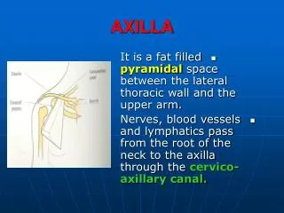
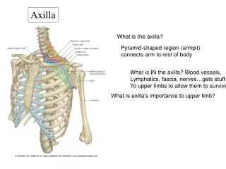
The Axilla • Is an irregular pyramidal shaped space between the upper part of the arm and the side of the chest through which major neurovascular structures pass between neck & thorax and upper limb. • Axilla has an apex, a base and four walls. Forming the clavi-pectoral canal
Boundaries of the Axilla • Apex: • Upper end of axilla or APEX is directed into the root of neck • Bounded in front by the clavicle • Behind by upper border of scapula • Medially by outer border of the 1st rib
cervico axillary canal • is bounded, by 3 bones: • Clavicle anteriorly. • Upper border of the scapula posteriorly. • Outer border of the first rib medially. • It is called cervico-axillary canal. 6
Base: • Formed by skin, superficial fasciae, & deep fascia • stretching between the anterior and posterior walls. • It is bounded: • In front by the anterior axillary fold (formed by the lower border of the Pectoralis major muscle). • behind by the posterior axillary fold (formed by the tendons of latissimusdorsi and teres major muscles). • medially by upper 4 to 5 ribs & the chest wall. • Lateral: the arm
The Deep fascia: or The axillary fascia extending between Pectoralis Major (ant fold) and Lat Dorsi & Teres Major (post fold) • Supported by Suspensorylig of Axilla • The clavi-pectoral fascia • It is a strong sheet of connective tissue • Split above to enclose the subclavius muscle and is attached to the clavicle • Below it splits to enclose the pectoralis minor muscle • Then continues downward as the suspensory ligament of the axilla • Then joins the fascial floor of armpit
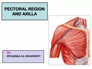
Anterior wall: Is formed by Pectoralis major * Pectoralis minor ** Subclavius Clavipectoral fascia ** *
Medial wall • Upper 4 ribs with intercostal muscles • Serratus ant (upper portion) with fascia • Long thoracic nerve deep to fascia • Intercostobrachial nerve pierce medial wall
Posterior wall: • Is formed by: • Subscapularis • Latissimusdorsi • Teres major muscles Subscapularis Teres major Lattisimus dorsi
Quadrangular space The inferior margin of the Subscapularis The lateral margin of the long head of the Triceps brachii The surgical neck of the Humerus The superior margin of the Teres major Passing through the quadrangular space are Axillary nerve Posterior circumflex humeral artery and vein.
Triangular space • the medial margin of the long head of the triceps brachii muscle; • the superior margin of the teres major muscle; • the inferior margin of the subscapularis muscle • Passing through the triangular space are • circumflex scapular artery and vein
Triangular interval The inferior margin of the teres major muscle Passing through the Triangular interval is the Radial nerve The lateral margin of the long head of the Triceps brachii The shaft of the humerus
Lateral wall • Bicipital groove of humerus with • Teres Major • Lattisimus dorsi • Pectoralis major • Long head of biceps • Short head of biceps • Coracobrachialis 18
The medial wall: • Is formed by: • Serratus anterior • Upper 4 or 5 ribs & Intercostal muscles . • The lateral wall: • Is formed by: • Coracobrachialis • Biceps brachii • Intertubercular groove of the humerus.
Deltoid muscle • posterior fibers of the deltoid – arm extension • Middle fibers of the deltoid – arm abduction
Contents of Axilla • Axillary artery and its branches • Axillaryvein and its tributaries • Lymph vessels and lymph nodes • Important nerve plexus the “Brachial Plexus” which innervates the upper limb • Cords and braches of brachial plexus. • Axillary artery and its branches. • Axillary vein and its tributaries. • Axillary lymph nodes. • Axillary fat. • Loose connective tissue.
Contents of The Axilla The neurovascular bundle is enclosed in connective tissue sheath, called ‘axillary sheath’ Axillary sheath is continuous with the prevertebral fascia
Axillary Artery • Is a continuation of subclavian artery • Begins at the lateral border of the 1st rib • Ends at the lower border of teres major • It continues as the brachial artery • Closely related to brachial plexus cords • Enclosed with them in the axillary sheath • Pectoralisminor divides it into 3 parts
The axillary artery is separated into three parts by the pectoralis minor muscle, which crosses anteriorly to the vessel. • the first part is proximal to pectoralis minor(extends from the lateral border of 1st rib to medial border of P.minor) • the second part is posterior to pectoralis minor (behind the P. minor) • the third part is distal to pectoralis minor (the longest part, extending from the lateral border of P.minor to the lower border of teres major muscle.
Branches • Generally, six branches arise from the axillary artery: one from the first part, two from the second part and three from the third part. • First part; Branches: Superior thoracic artery, a small branch supplying first intercostal space. Relations: • Anterior: Pectoralis major, covering fascia, skin, cephalic vein • Posterior: Long thoracic nerve • Lateral: Three cords of brachial plexus • Medial: Axillary vein
Second part; • a. The thoraco-acromialbranch which pieces the clavipectorial fascia and divides into four branches thus; • The deltoid branch which lies in delto-pectoral groove. • The clavicular branch which supplies sternoclavicular joint and subclavius muscle • The pectoral branch which supplies the pectoral muscles • The acromion branch which takes part in the anastomosis over the acromial process b. The lateral thoracic artery, which runs along the lateral border of the P. minor muscle and supplies the anterior and medial walls. In females branches emerge from the inferior border of the P. major and contribute in the supply of the breast.
Relation • Anterior: Pectoralis minor and major, covering fascia and skin • Posterior: Posterior cord of brachial plexus • Lateral: Lateral cord of brachial plexus • Medial: medial cord of brachial plexus and axillary vein
Branches of the Third part; • a) The anterior circumflex humeral artery, It passes anterior to the surgical neck of the humerus and anastomoses with the posterior circumflex humeral artery. • Supplies branches to surrounding tissues, which include the glenohumeral joint and the head of the humerus. • b) The posterior circumflex humeral artery; • A larger artery than the anterior and accompanies the axillary nerve through the quadrangular space. • Supplies the glenohumeral joint and surrounding muscles i.eteres major, minor and long head of triceps brachii.
Relation • Anterior: Pectoralis major, medial root of the median nerve • Medial: Ulnar nerve, axillary vein, medial cutaneous nerve of the arm • Posterior: subscapularis, latissimus dorsi and teres major • Lateral: Coracobrachialis, biceps, humerus
AXILLARY VEIN • The axillary vein begins at the lower margin of the Teres major muscle and is the continuation of the basilic vein, which is a superficial vein that drains the posteromedial surface of the hand and forearm and penetrates the deep fascia in the middle of the arm. • The axillary vein passes through the axilla MEDIAL AND ANTERIOR TO THE AXILLARY ARTERY and becomes the subclavian vein as the vessel crosses the lateral border of 1st rib at the axillary inlet. • Tributaries of the axillary vein generally follow the branches of the axillary artery. Other tributaries include Brachial veins that follow the brachial artery, and the Cephalic vein.
The cephalic vein is a superficial vein that drains the lateral and posterior parts of the hand, the forearm, and the arm. • In the area of the shoulder, it passes into an inverted triangular cleft (the clavipectoral triangle) between the deltoid muscle, pectoralis major muscle, and the clavicle. • In the superior part of the clavipectoral triangle, the cephalic vein passes deep to the clavicular head of the pectoralis major muscle and pierces the clavipectoral fascia to join the axillary vein. • Many patients who are critically unwell have lost blood or fluid, which requires replacement. Access to a peripheral vein is necessary to replace the fluid. The typical sites for venous access are the cephalic vein adjacent to the anatomical snuffbox or the antecubital veins, which lie within the superficial tissues of the cubital fossa.
Axillary Lymph Nodes • The fibro-fatty connective tissue of the axilla has many lymph nodes. • They are arranged in five principal groups: apical, pectoral, subscapular, humeral, and central.
Axillary Lymph Nodes • Anterior or Pectoral group receive lymph from upper half of anterior wall trunk and from major part of breast. • Posterior or Scapular group receive lymph from posterior wall of upper half of trunk and from axillary tail of breast. • Lateral group receives lymph from upper limb. • Central group receives lymph from preceding groups and drains into apical group. (intercostobrachial N) • Apical or infraclavicular (subclavian) group lie deep to clavipectoral fascia. They receive lymph from the central group, from upper part of breast and from the thumb.
What is a Brachial Plexus ? Brachial Plexus is a network of nerves present at the root of the neck to enter the upper limb . It is formed by the union of the anterior Rami of the C 5th, 6th, 7th & 8th and the 1st thoracic spinal nerve. Brachial Plexus supplies muscles and skin of upper limb except trapezius supplied by spinal accessory nerve and an area of skin of axilla supplied by intercostobrachial nerve 39
Organization of brachial plexus • 5 Roots unite into 3 trunks in the neck: • Roots of C5 & C6 unite - Upper trunk • Root of C7 continues ---Middle trunk • Roots of C8 & T1 unite - Lower trunk • Divisions: Each trunk divides into anterior(flexor)and a posterior(extensor) division • Cords • Anterior divisions of the superior and middle trunks unite to form the lateral cord. • Anterior division of the inferior trunk form the medial cord. • Posterior divisions of all 3 trunks unite to form the posterior cord. Important • The roots lie between scalene muscles. • The trunks in the posterior triangle. • The divisions are behind the clavicle. • The cords and branches are situated in axilla
Supraclavicular branches (4) • The dorsal scapular nerve (C5), posterior to the roots –Supplies rhomboids major, rhomboid minor and levator scapulae. • The nerve to subclavius (C5, 6), anterior to the roots –supplies subclavius • The long thoracic nerve (C5, 6, 7) posterior to the roots-supplies serratus anterior • The suprascapular nerve (C5, 6) –supplies supraspinatus and infraspinatus.
Branches of Lateral Cord • lateral pectoral nerve (C5, 6, 7) pectoralis major and minor • musculocutaneous nerve (C5, 6,7)coracobrachiais, brachialis and bicep brachii. • lateral root of the median nerve (C5, 6, 7) Branches of Medial cord • Medial pectoral nerve (C8, T1) • Medial root of the median nerve (C8, T1) • Ulnar nerve (C7, 8, T1) • Medial cutaneous nerve of the arm (or medial brachial cutaneous nerve; C8, T1) • Medial cutaneous nerve of the forearm (or medial antebrachialcutaneous nerve; C8, T1) .
Branches of Posterior cord • The Upper subscapular nerve (C5, 6) –supplies subscapularis muscle. • Thoracodorsal nerve (C5, 6, 7) –supplies latissimus dorsi • Lower subscapular nerve (C5, 6)-supplies subscapularis and teres major. • Axillary nerve (C5, 6)-supplies deltoid and teres minor • Radial nerve (C5, 6, 7, 8, T1)-nerve of extensor compartment of arm and forearm.
Intercostobrachial nerve • The lateral cutaneous branch of the second intercostal nerve does it is named the intercostobrachial nerve. • It pierces the external intercostal and Serratus anterior, crosses the axilla to the medial side of the arm, and joins with a filament from the medial brachial cutaneous nerve. • It supplies the skin of the upper half of the medial and posterior part of the arm. • The intercostabrachial nerve is also sometimes divided in axillary node clearance.
Apical group; consists of lymph nodes at the apex of the axilla. • Located along the medial side of the axillary vein and the first part of the axillary artery. • It receives lymph from all other groups of axillary lymph nodes. • Pectoral (anterior) group; • Consists of three to five lymph nodes that lie along the medial wall of the axilla, around the lateral thoracic vein and the inferior border of the pectoralis minor. • The pectoral group of nodes receives lymph mainly from the anterior thoracic wall including the breast. • The subscapular (posterior) group; • Consists of six or seven lymph nodes that lie along the posterior axillary fold and subscapular blood vessels. • This group of lymph nodes receives lymph from the posterior aspect of the thoracic wall and scapular region.
The humeral (lateral) group; • Consists of four to six lymph nodes that lie along the lateral wall of the axilla, medial and posterior to the axillary vein. • This group of lymph nodes receives nearly all the lymph from the upper limb, except that carried by lymphatic vessels accompanying the cephalic vein, which drains to the central and apical axillary nodes. • Central group; • The central group of axillary lymph nodes consists of three or four large lymph nodes situated deep to the pectoralis minor near the base of the axilla, in association with the second part of the axillary artery. • As its name indicates, the central group receives lymph from the pectoral, subscapular, and humeral groups of axillary lymph nodes.