Download
1 / 55
550 likes | 560 Views
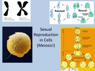
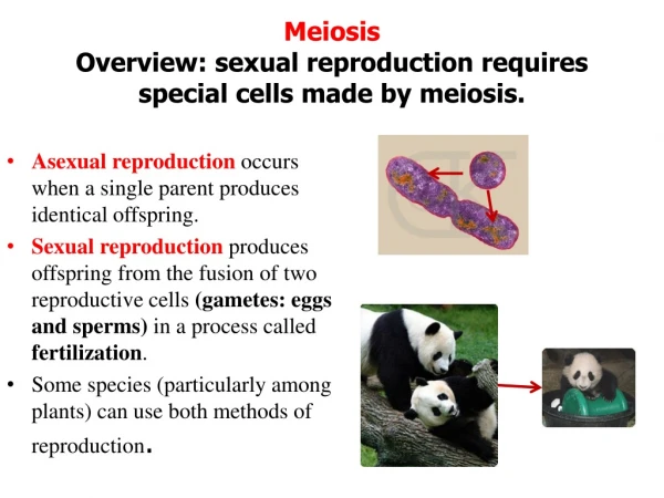
Delve into the intricate processes of spermatogenesis, ovogenesis, and fertilization in human reproductive biology. Explore the journey of gametes, from development to union, in creating new life.

E N D
THE SEXUAL CELLSEMBRIOGENESIS ASSOCIATE PROFESSOR IOLANDA BLIDARU MD,PhD
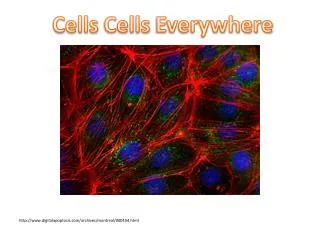
The sexual cells = gametes The spermatozoon • Origin - spermatogenesis • seminiferous tubuleepithelium of the testis • - fibromuscular wall • - Sertoli cells • - germcellsin different stages • - vessels • - Leydigcells(steroidogenesis)
The spermatozoon Spermatogenesis: 74 days / continuously Spermatogonia (46XY) ▼ mitosis Spermatocyte I (46XY) ▼meiosis Spermatocytes II (23X) + (23Y) ▼ mitosis Spermatides (23X) + (23Y) + (23X) + (23Y) ▼metamorphosis Spermatozoa (4)
The spermatozoon morphology The head • The acrosome: hydrolytic enzymes (hyaluronidase, acrosine) mecanisms of fertilization The connective piece The flagellum (the tail) • microtubular complex • sliding motility (wave 180° rotation wave)
The pre-fertilization transformations of the spermatozoa • Motility • Fertilization capability • Capacitation – in the female genitaltract • increased motility • loosing material from the acrosome surface • exposing the receptors • The acrosome reaction - a final maturation
The passage of thespermatozoain the female genital tract The vagina • pH = 5 (semen- pH = 7) • 5 min – 1 hour (the vagina → the tube) The cervical canal • filter & reservoir (200.000 – 400.000, 24 hours) • cervical mucus (tricot-like) – permisivity The utero-tubal junction • filter & reservoir • constant concentration (1000 → a few hundreds in the ampullary part of the tube, 2-34 hours)
The ovum Ovogenesis & Folliculogenesis The embrionic-fetal life germinal epithelium (the cords) = primordialfollicles Theprimordialfollicle • ovogonia 20 microns ▼ mitosis • oocyte I ▼ blocked in the prophase of the first meiotic division ! • granulosa cells layer • basal membrane Slavjanski
Ovogenesis & Folliculogenesis At puberty- 300.000 follicles in the ovaries Folliculogenesis follicle maturation - 3 months The primary follicle • oocyte I (30-60 microns) • granulosa cells layer • zonapellucida • Slavjanski membrane
Ovogenesis & Folliculogenesis The secondary follicle • oocyte I(45-70 microns) The tertiary follicle (Call – Exner follicle) • oocyte I(60-80 microns) • granulosa cell massif • zona pellucida • theca interna (cellular) • theca externa (fibrilar) The antral follicle • oocyte I(90 microns) + • cumulus proliger – cAMP • corona radiata
Ovogenesis & Folliculogenesis The mature follicle (de Graaf)15-20mm, unique/cycle • oocyte I(100 microns), peripheral • granulosa membrane • Slavjianski membrane • theca interna E • theca externa • follicular cavity -follicularfluid • 15-50 years → 13 ovulations / year
Folliculogenesis The follicular development • recruitment • selection • dominance
Ovogenesis & Folliculogenesis • The dominant follicle → increased 17β estradiol (8-th day) → atresiaof the rest of follicles (both ovaries) → LH & FSH peak → ovulation • Before the ovulation – meiosis restarts ▼ oocyte II (22x) + the first polar body
Ovulation 24 h 16-40 h • E2 peak LH peak ovulation The ovumcan be fertilized 24 ore post-ovulation • The effectsof the LH peak • the continuation of the meiosis • the release of thefirst polar body • OMI inhibition (OMI cumulus cells meiosis inhibition ←cAMP) • luteinization • ovumrelease
Ovulation The process of ovum transfer from the ovary to the place of fertilization. • The phenomena: complex • Follicular apex → pellucida membrane rupture → stigmaformation → ovulation(oocyte II + cumulus + granulosa cells + follicular fluid release) → grasped by the fimbriated extremity of the tube • The granulosa and theca interna cells → luteal cells (corpus luteum)
Fecundation Fertilization = a diploid egg = zygote • The ovumtransferfrom the ovary to the external ⅓ of the tube- 3 mecanisms • intra-tubal negative pressure • the contraction of the tubal fimbria • the contact between fimbriatedtubal extremity and the cumulus • The spermatozoatransferin the external ⅓ of the tube • The granulosa cells(cumulus oophorus) resorbtion
Fecundation 4. The sperm interactionwith zona pellucida andits penetration 5. Transformation of the sperm head into male pronucleus 6. Transformation of the ovum nucleus into female pronucleus (release of the 2-nd polar globe) + pronuclei attachment 7.Cromosome union ► the zygote (44+XX / XY)
Transformation of the head into male pronucleus. Transformation of the ovum nucleus into female pronucleus (release of the 2-nd polar globe) + pronuclei attachment
Segmentation. Migration • Tubal migration + segmentation (cleavage) (3-4 days) ↓ 4 blastomeres (four cell stage) ↓ 8 blastomeres (eight cell stage) ↓ unequal division • micromeres (small, clear, outer mass ► trophoblast) • macromeres (large, dark cells ► embryo) ↓ morula (12-16 blastomeres, fine zona pellucida) ↓ blastocist cavity fomation (ZP disappears) macromeres embryo button
Segmentation. Migration Migration (tubal transport) • muscle contractility • epithelial cilia activity • tubal fluid
Implantation Post-conceptional – 7 days – up to the implantation - 3 days – the egg is in uterus The stages of implantation • Preimplantation • Attachment (apposition) – to the endometrium • Nidation – the blastocyst penetrates into the endometrium → decidual transformation • Placentation – a connection between the endometrial vessels and the trofoblastic lacunae
Implantation 1. The preimplantation stage • the apical membranes are not in contact • the blastocyst → nurturition by “grasping” mechanism 2. The attachment stage • zona pellucida dettachment in the day 6 • membrane attachment day 6-7 • syncronization of the blastocyst - endometrium alterations the endocrine profile of the implantation
Implantation The 1-st week • superficial blastocyst implantatationat the fundus • abnormal implantation ectopic pregnancy, placenta praevia The 2-nd week • Fulfilling of the implantation • Enlargment of the contact with endometrium • trophoblast diferentiation Endometrium decidua (caduca)
Implantation The decidua consists of three layers: • The superficial compact layer - decidual cells • The spongy (deep) layer - with glands • The thin basal layer. • The separation of placenta occurs through the spongy layer • While the endometrium regenerates again from the basal layer.
Implantation. Development of the egg 2-nd Week - embryonic button 2 layers = embryonicdisc (endoderm + ectoderm) - amnionic cavity(between endoderm + ectoderm) - Heuser membrane delimits primitive yolk sac lecytocel - lacunae in syncytiotrophoblast) - embryotroph fluid diffusion embryonic disc maternal blood (endometrial capillaries) + eroded glands secretions
Implantation. Development of the egg Day 10 – 2-nd week • the egg is completely included inside the endometrium (protrudes) • onset of the utero-placental circulation = opening of the uterine vessels into ST lacunae – fusion = network – intervillous space • up to the end of week 2 – proliferation of CT inside ST the solid primitive villi