Download

1 / 38
420 likes | 640 Views
Fundamental Nursing Chapter 30 Urinary Elimination.

E N D
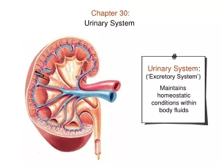
The urinary system (Fig. 30-1) consists of the kidneys, ureters, bladder, and urethra. These major components, along with some accessory structures such as the ring-shaped muscles called the internal and external sphincters, work together to produce urine (fluid within the bladder), collect it, and excrete it from the body.
Urinary elimination (the process of releasing excess fluid and metabolic wastes), or urination, occurs when urine is excreted. Under normal conditions, the average person eliminates approximately 1500 to 3000 mL of urine each day. The consequences of impaired urinary elimination can be life-threatening.
The need to urinate becomes apparent when the bladder distends with approximately 150 to 300 mL of urine. The distention with urine causes increased fluid pressure, stimulating stretch receptors in the bladder wall and creating a desire to empty it of urine.
Patterns of urinary elimination depend on • physiologic • emotional, • social factors. • degree of neuromuscular development • and integrity of the spinal cord; • the volume of fluid intake and • the amount of fluid losses, • the amount and type of food consumed; • the person's circadian rhythm, • habits, • opportunities for urination, • and anxiety.
General measures to promote urination include: • providing privacy, • assuming a natural position for urination (sitting for women, standing for men), • maintaining an adequate fluid intake, and • using stimuli such as running water from a tap to initiate voiding.
Urine Specimen Collection Voided Specimens • A voided specimen is a sample of fresh urine collected in a clean container. The first voided specimen of the day is preferred because it is most likely to contain substantial urinary components that have accumulated during the night.
Clean-Catch Specimens • A clean-catch specimen is a voided sample of urine considered sterile and is sometimes called a mid-stream specimen because of how it is collected. • As soon as the specimen is collected, it is labeled and taken to the laboratory.
Catheter Specimens • A urine specimen can be collected under sterile conditions using a catheter, the nurse can aspirate a sample through the lumen of a latex catheter or from a self-sealing port (Fig. 30-2).
24-Hour Specimens • The nurse collects, labels, and delivers a 24-hour specimen (collection of all urine produced in a full 24-hour period) to the laboratory for analysis.
Abnormal Urine Characteristics • Hematuria: urine containing blood • Pyuria: urine containing pus • Proteinuria: urine containing plasma proteins • Albuminuria: urine containing albumin, a plasma protein • Glycosuria: urine containing glucose • Ketonuria: urine containing ketones
Abnormal Urinary Elimination Patterns • Anuria • Anuria means absence of urine or a volume of 100 mL or less in 24 hours. It indicates that the kidneys are not forming sufficient urine. • Oliguria • Oliguria, urine output less than 400 mL per 24 hours, indicates inadequate elimination of urine. • Residual urine, or more than 50 mL of urine that remains in the bladder after voiding
Polyuria • Polyuria means greater than normal urinary volume and may accompany minor dietary variations. For example, consuming higher than normal amounts of fluids, especially those with mild diuretic effects (e.g., coffee, tea), or taking certain medications actually can increase urination.
Nocturia • Nocturia (nighttime urination) is unusual because the rate of urine production is normally reduced at night. • Dysuria • Dysuria is difficult or uncomfortable voiding and a common symptom of trauma to the urethra or a bladder infection. Frequency (need to urinate often) and urgency (strong feeling that urine must be eliminated quickly) often accompany dysuria.
Incontinence • Incontinence means the inability to control either urinary or bowel elimination and is abnormal after a person is toilet-trained.
Assisting Clients with Urinary Elimination • Commode • A commode (chair with an opening in the seat under which a receptacle is placed) is located beside or near the bed (Fig. 30-3).
Urinal • A urinal is a cylindrical container for collecting urine. It is more easily used by males.
Using a Bedpan • A bedpan (seatlike container for elimination) is used to collect urine or stool. Figure 30-5 • Two types of bedpans: fracture pan (left) and conventional bedpan (right).
Catheterization • Catheterization (act of applying or inserting a hollow tube), A urinary catheter is used for various reasons: • Keeping incontinent clients dry (catheterization is a last resort that is used only when all other continence measures have been exhausted) • Relieving bladder distention when clients cannot void
Assessing fluid balance accurately • Keeping the bladder from becoming distended during procedures such as surgery • Measuring the residual urine • Obtaining sterile urine specimens • Instilling medication within the bladder
Types of Catheters • External Catheters • An external catheter (urine-collecting device applied to the skin) is not inserted within the bladder; instead, it surrounds the urinary meatus. Examples of external catheters are a condom catheter (Fig. 30-7)External catheters are more effective for male clients.
Figure 30-7 • A condom catheter is an example of an external urine collection device.
Straight Catheters • A straight catheter is a urine drainage tube inserted but not left in place. It drains urine temporarily or provides a sterile urine specimen (Fig. 30-9). • Retention Catheters • A retention catheter, also called an indwelling catheter, is left in place for a period of time (see Fig. 30-9). The most common type is a Foley catheter.
Figure 30-9 • Types of urinary catheters A) Retention (Foley) catheter with balloon. (B) Straight catheter.
Unlike straight catheters, retention catheters are secured with a balloon that is inflated once the distal tip is within the bladder. Both straight and retention catheters are available in various diameters, sized according to the French scale: for adults, sizes 14, 16, and 18 F are commonly used.
Providing Catheter Care • “Catheters left in place for more than a few weeks become encrusted or obstructed, and lead to infection. In addition, bacteria that adhere to the urinary catheter develop a complex biologic structure, which protects them from antibiotics”
Catheter care (hygiene measures used to keep the meatus and adjacent area of the catheter clean) helps to prevent the growth and spread of colonizing pathogens.
Figure 30-11 • Techniques for suspending a drainage system below the bladder: ( A) wheelchair patient; (B) ambulating patient with and without an IV pole.
Continuous Irrigation • A continuous irrigation (ongoing instillation of solution) instills irrigating solution into a catheter by gravity over a period of days (Fig. 30-12). • Continuous irrigations keep a catheter patent after prostate or other urologic surgery in which blood clots and tissue debris collect within the bladder.
Figure 30-12 • Bladder irrigation using a three-way catheter
Urinary Diversions • In a urinary diversion, one or both ureters are surgically implanted elsewhere. This procedure is done for various life-threatening conditions. The ureters may be brought to and through the skin of the abdomen. A urostomy (urinary diversion that discharges urine from an opening on the abdomen), or implanted within the bowel (called an ileal conduit) (Fig. 30-14). • SPC
Figure 30-14 • Examples of urinary diversions. (A) Ileal conduit. (B) Cutaneous ureterostomy.
Nursing Implications • Self-Care Deficit: Toileting • Impaired Urinary Elimination • Risk for Infection • Stress Urinary Incontinence • Urge Urinary Incontinence • Reflex Urinary Incontinence • Total Urinary Incontinence • Functional Urinary Incontinence • Situational Low Self-Esteem • Risk for Impaired Skin Integrity