Spinal Cord
Spinal Cord. Dr Rania Gabr. Objectives. Describe the gross anatomical features of the spinal cord. Describe the level of the different spinal segments comparing to the level of their respective vertebrae. Identify important gross features of spinal cord, nerve roots, and spinal ganglia.


Spinal Cord
E N D
Presentation Transcript
Spinal Cord Dr Rania Gabr
Objectives • Describe the gross anatomical features of the spinal cord. • Describe the level of the different spinal segments comparing to the level of their respective vertebrae. • Identify important gross features of spinal cord, nerve roots, and spinal ganglia. • Describe the internal features of spinal cord (gray matter and white matter) in the different regions.
Vertebral Column • Consists of vertebrae and the intervening discs. • It appears as straight line anteroposteriorly, and with multiple curves laterally • According to shape and location, vertebrae can be divided into: • Cervical (7) • Thoracic (12) • Lumbar (5) • Sacral (5 Fused ) • Coccyx ( 4 Fused)
Nervous system Central nervous system • Brain • Spinal cord Peripheral nervous system • 12 pairs of cranial nerves • 31 pairs of spinal nerves
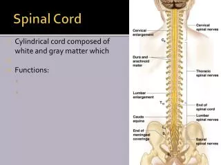
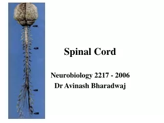
Structure of the Spinal Cord • A cylinder of gray & white matter • In the upper 2/3 of vertebral canal • Extends From foramen magnum to L1 (or L2) • Covered with meninges & CSF • A typical adult spinal cord ranges between 42 and 45 centimeters .
The Spinal Cord • The spinal cord is associated with 31 pairs of spinal nerves that connect the CNS to muscles, receptors, and glands. • Each side of the spinal cord contains : • 8 Cervical nerves (C1-C8) • 12 Thoracic nerves (T1-T12) • 5 Lumbarnerves (L1–L5) • 5 Sacral nerves (S1–S5), • 1 Coccygealnerve
Structure of the Spinal Cord • The spinal cord is shorter than the vertebral canal that houses it.Extends only to L1/ L2 • Has two enlargements, cervical & lumbar due to cells and fibers of limbs. • Ends inferiorly in a tapering conusmedullaris • Anchored to the coccyx by a meningeal ( non neuronal) extension (filumterminale) • Held to dura by denticulate ligament
The cervical enlargement consists of cord segments C3-T1and provides innervation for the upper limb via the brachial plexus. • The lumbar enlargementis made up of segments L1-S3 and is associated with innervation of the lower limb via the lumbar plexus (L1-L4) and the sacral plexus (L4-S2)
The Spinal Cord Provides a vital link between the brain and the rest of the body. Exhibits some functional independence from the brain. The spinal cord and its attached spinal nerves serve some important functions :-
Functions of the Spinal Cord • Receives afferent fibers from sensory receptors of the trunk and limbs • Controls movements of the trunk and limbs • Provide autonomic innervation for most of the viscera. • Responsible for many loop reflexes • It conveys afferent information to higher centers and mediate their controlling influence over spinal mechanisms.
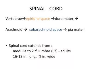
Spinal Meninges • Are continuous with the cranial meninges. • Structures that encircle the spinal cord, listed from outermost to innermost are: 1-vertebra 2-epidural space: Contains blood vessels, areolar connective tissue and fat. 3-dura mater 4-subdural space: a potential cavity between the dura and arachnoid mater, contains a small volume of serous fluid 5-arachnoid 6-subarachnoid space:Contains cerebrospinal fluid (CSF) and blood vessels 7-pia mater
Surfaces • Surface marked by furrows • A deep ventral (anterior) median fissure • A dorsal (posterior) median sulcus • 2 dorsolateral & 2 ventrolateral sulci • 2 dorsal intermediate sulci
Cross Section of Spinal Cord • Anterior median fissure and posterior median sulcus • deep clefts partially separating left and right halves • Gray matter: neuron cell bodies, dendrites, unmylinatedaxons • Divided into horns • Posterior (dorsal) horn • Anterior (ventral) horn • Lateral horn only in thoracic, lumbar and sacral. The intermediate zone traversed by Central Canal.
White matter • Myelinated axons • Divided into threecolumns(funiculi) • Ventral • Dorsal • lateral • Each of these divided into sensory or motortracts • Commissures: connections between left and right halves • Gray with central canal in the center • White • Roots • Spinal nerves arise as rootlets then combine to form dorsal and ventral roots • Dorsal and ventral roots merge laterallyand form the spinal nerve
Grey matter • Consists of nerve cell bodies and their processes, neuroglia, and blood vessels • The nerve cells are multipolar and are of three main categories: • Sensory neurons (Tract cells), which receive impulses from the periphery of the body and whose axons constitute the ascending fasciculi of the white matter, are located in the dorsal horns • Lower motor neurons, which transmit impulses to the skeletal muscles, are located in the ventral horns (similar neurons in the lateral horn are the preganglionic neurons of the autonomic system) • Interneurons (connector neurons) : linking sensory and motor neurons, at the same or different levels, which form spinal reflex arcs.
Different cord levels vary in the relative amounts and configuration of grey and white matter. • Higher levels contain greater amounts of white matter. • Why? • Because ascending tracts gain fibresat each successive level, whereas the opposite is true of descending tracts.
The size and shape of the dorsal and ventral horns varies according to the level. • Both dorsal and ventral horns are, therefore, particularly well developed at cervical and lumbar levels in association with innervation of the upper and lower limbs.