Download
1 / 41
550 likes | 1.57k Views
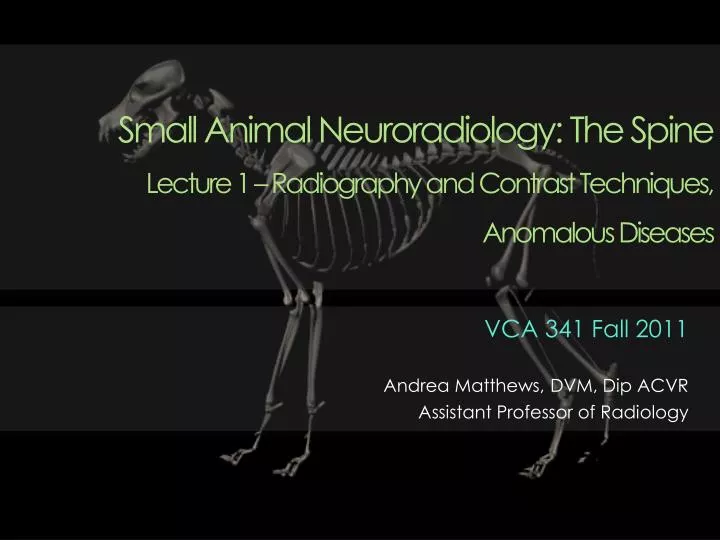
Small Animal Neuroradiology: The Spine Lecture 1 – Radiography and Contrast Techniques, Anomalous Diseases . VCA 341 Fall 2011 Andrea Matthews, DVM, Dip ACVR Assistant Professor of Radiology. Normal Anatomy. Canine and feline vertebral formulas. Normal Anatomy – Cervical.

E N D
Small Animal Neuroradiology: The SpineLecture 1 – Radiography and Contrast Techniques, Anomalous Diseases VCA 341 Fall 2011 Andrea Matthews, DVM, Dip ACVR Assistant Professor of Radiology
Normal Anatomy Canine and feline vertebral formulas Matthews
Normal Anatomy – Cervical C1 (or “atlas”) • Central arch and two wide horizontal wings perforated by transverse foramina C2 (or “axis”) • Long, thin spinous process which overlaps the dorsal arch of C1 • Odontoid process (dens) C6 • Expanded transverse process ventrally Matthews
Cervical Spine C1 C2 C3 C5 Matthews
Cervical Spine atlas C1 axis C2 C3 C5 Matthews TUSCVM
Normal Anatomy – Thoracic Rib heads articulate with cranial aspect of corresponding vertebral bodies Spinous processes change direction from caudal angulation to cranial angulation at the anticlinal vertebra(usually T11) Accessory processes on last 4-5 thoracic vertebrae T10-11 intervertebral disc space is normally narrow Matthews
Thoracic Spine Anticlinal vertebra Anticlinal disc space is narrow normally T11 Proximal ribs T10 TUSCVM Matthews
Normal Anatomy - Lumbar Lumbar vertebral bodies are longer than thoracic vertebrae • Especially in cats. Transverse processes are angled cranially, laterally and somewhat ventrally Accessory processes (present on the first four vertebrae) can be especially large in cats “Fuzzy” ventral margin of L3 and L4 • Due to attachment of the diaphragmatic crura (especially in large dogs). } Do not mistake for mineralized intervertebral disc material! Matthews
Lumbar Spine TUSCVM Matthews
Thoracic Spine Accessory process TUSCVM Matthews
Lumbar Spine Attachment for diaphragmatic crus L3 L4 TUSCVM Matthews
Sacral / Caudal Vertebra Sacrum • Lumbosacral angulation can vary significantly between individuals • Changes with degree of flexion or extension Caudal Vertebra • Formerly known as coccygeal vertebrae • Vary in number • Hemal arches ventrally Matthews
Sacral / Caudal Vertebra Matthews
Lumbosacral Junction Ilial wings Articular facet joint TUSCVM Matthews
Lumbosacral Junction Margins of the sacrum Matthews TUCSVM
Typical Vertebra Matthews
Ligamentous Structures Konig and Liebich, Veterinary Anatomy of Domestic Animals, 3rd Ed Matthews
Survey Radiography Lateral and ventrodorsal views Adequate relaxation is required for good positioning • General anesthesia preferred • Exception: Suspected fracture and/or luxation • Can obtain lateral and horizontal beam Collimation to improve quality Matthews
Survey Radiography Lateral cervical radiograph Ventrodorsal cervical radiograph Matthews
Survey Radiography Lateral thoracic radiograph Matthews
Survey Radiography Beware of “pseudonarrowing” of disc spaces • Artifactual narrowing due to divergence of x-rays Kishigami, Y.et al. Vet Radiol Ultrasound 41, 9–18 (2000). Matthews
Pseudonarrowing Matthews
Radiography and Contrast Techniques Matthews
Myelography Introduction of contrast into subarachnoid space • Water soluble, iodinated, non-ionic contrast media Sites of injection • Cisterna magna • More likely to seizure • Difficult to get flow caudally in some cases • Lumbar (L5-6, L4-5) • Possible epidural leakage • More difficult technically • Fewer complications • Better flow of contrast typically Matthews
Myelography Site for cervical injection Site for lumbar injection Diaz, F. In Practice 27, 502-510 (2005). Matthews
Myelography • Contraindications • Inflammatory disease (meningitis) • Bleeding diatheses • Evidence of vertebral instability (could increase spinal cord damage) Indications • Neurologic signs with no lesion on survey rads • Multiple lesions seen on survey rads • Single lesion seen on survey rads not consistent with clinical signs • Abnormality on survey rads which needs further characterization Matthews
Normal Myelogram Courtesy Dr. L. Pack Matthews
Extradural Lesion Extruded intervertebral disc material L3 L3 L4 Matthews
Intradural/Extramedullary Lesion Golf tee sign Matthews
Intramedullary Lesion Widening of spinal cord due to spinal cord tumor (glioma) Courtesy Dr. L. Pack Matthews
Anomalous Diseases Matthews
Hemivertebra Failure of vertebral body to develop fully • Persistence of sagittal membrane (notochord) Most commonly in thoracic spine • May have focal kyphosis Often incidental finding Bulldogs, Boston terrier and pugs (“screw-tailed” breeds) Matthews
Hemivertebra Butterfly vertebra Matthews TUCSVM
Block Vertebra Fusion of two or more adjacent vertebrae • Involve bodies, laminae and pedicles or entire vertebrae • Incomplete development of intervertebral disc Can occur at any location in spine Incidental • Differentiated from healing fractures, luxations, discospondylitis Matthews
Block Vertebra TUCSVM Matthews
Transitional Vertebra Vertebra at the junction between two spinal regions that assumes the characteristics of both regions • Thoracolumbar, lumbosacral and sacrocaudal junctions Usually incidental findings • Important when identifying surgical site • Make positioning of VD pelvis difficult • Can be associated with lumbosacral instability Matthews
Transitional Vertebra Sacralization of L7 Matthews
Spina Bifida Part of general defect called Spinal Dysraphism • Failure of neural arch to close during embryogenesis Two types • Spina bifida occulta • No spinal cord or meningeal involvement • No clinical signs typically • Spina bifida manifesta • Protrusion of the meninges (meningocele) or meninges and spinal cord (meningomyelocele) • Associated clinical signs Matthews
Anomalous Diseases Failure of fusion of spinous processes Matthews • Common in screw-tailed breeds • Bulldogs, Boston Terriers, Pugs, Manx cats
Other Anomalies Scoliosis • Lateral bowing as seen on a VD or DV view Lordosis • Ventral bowing as seen on a lateral view Kyphosis • Dorsal bowing as seen on a lateral view Scoliosis (from Radiographic Interpretation for the Small Animal Clinician 2nd Ed) Matthews
The End Matthews