Download
1 / 13
130 likes | 269 Views
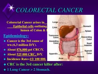
Cancer Care Engineering Colorectal Cancer Gabriela Chiorean, M.D. June 26, 2009. Rationale in colorectal cancer. Perform OMICs of healthy, polyps, cancer Compare OMICs between cancer, polyps and healthy: develop new screening and risk assessment tools

E N D
Cancer Care Engineering Colorectal CancerGabriela Chiorean, M.D. June 26, 2009
Rationale in colorectal cancer • Perform OMICs of healthy, polyps, cancer • Compare OMICs between cancer, polyps and healthy: develop new screening and risk assessment tools • Analyse changes in OMICs with treatment and correlate with response/toxicity: predictive markers • Mathematical modeling and bio-mapping • Cancer care delivery
Rationale: CCE now GENOMICS METABOLOMICS GLYCO- PROTEOMICS LIPIDOMICS C R C BIOMAP Mathematical modeling
Schema IUCRO-0221CCE in CRC active April 2009 S A M P L E S Blood (Serum) 7 mL red top Metab, vit D S H I P D R Y I C E • N=270 • Stratification: • Healthy (n=90) • Polyps (n=90) • Cancer (n=90) • stg 1/2 • stg 3 • stg 4 metastatic Blood (Plasma) 21 mL purple top Genomics, lipidomics, glycoproteomics Tissue 10 mg polyp or 50 mg cancer / 50 mg normal tissue 8-hr fasting
N= 5 Samples CollectionHealthy Controls Sign ICS (RN) Label specimens Healthy if no polyps/tumor Screening Colonoscopy – GI Clinic Collect by RN/processing CRS Blood 1x 7 mL glass red top 3 x 7 mL plastic lavender Questionnaires diet/environmental exposures
N= 3 Samples CollectionAdenomatous Polyps Sign ICS (RN) Polyps identified Label specimens Polyp Screening Colonoscopy – GI Clinic Collect by RN/processing CRS Blood 1x 7 mL glass red top 3 x 7 mL plastic lavender Tissue procurement/Research specialist -Polyp cut in ½ -Place in tube with no preservative -Freeze at -70oC Questionnaires diet/environmental exposures
Samples CollectionCancer N= 8 Call tissue procurement -Tumor tissue ~ 50 mg -Normal mucosa ~ 50 mg -Place in tube with no preservative -Freeze at -70oC Sign ICS (RN) Chemotherapy Follow-up Surgery Collect by RN/processing tissue procurement Blood: 1 x 7 mL red top glass tube 3 x 7 mL lavender plastic tubes Questionnaires: diet/environmental exposures Every 3 months Up to 24 months
CCE Blood Acquisition Protocols Glass Red Top Tube (1) Volume = 7mL Glass Purple Top Tubes (EDTA) (3) Volume = 7mL /tube Following blooddraw, patients and care givers administered diet and life style questionnaire Page: Amber Allen (page #) for transport to laboratory (RT) and processing 0.2 mL (2) Whole Blood into freezing tubes containing comet assay solution, mix, place on dry ice, FREEZE (-80oC) Maximum time at RT from draw to centrifugation: 45-60 min. Centrifuge: 1500g, RT, 15 min Maximum time at RT from draw to Whole Blood Removal: 20 min. Remaining whole blood Serum ( ~ 3mL), place on wet ice Maximum time at RT from draw to centrifuge: 30 min. Centrifuge: 1750g, RT, 15 min REGULAR EPPENDORF TUBES 0.3 mL (2) FREEZE (-80 oC) Metabolomics NMR 0.2 mL (2) FREEZE (-80 oC) Metabolomics MS 0.5 mL (2) FREEZE (-80 oC) Vitamin D Analysis Plasma (~ 6mL), place on wet ice Pellets (2); resuspend (1), combine with second pellet, re-centrifuge 1750g RT 5 min, decant, place on dry ice: FREEZE (-80 oC) SNP SILICONIZED EPPENDORFTUBES 0.2 mL (2) FREEZE (-80 oC) Lipidomics REGULAR EPPENDORF TUBES 1.5 mL (1) FREEZE (-80 oC)Glycoproteomics 0.2 mL (1) FREEZE (-80 oC) Proteomics 1.5 mL (1) LONG TERM STORAGE (LIQUID N2); Regular Eppendorf Tubes 0.2 mL (12) LONG TERM STORAGE (LIQUID N2); Siliconized Eppendorf Tubes 0.5 mL (2) LONG TERM STORAGE (LIQUID N2)
Metabolomics Typical 2D GCxGC/MS data from a colon cancer patient serum sample. After derivitization, approximately 800 metabolites are observed (many of the lower intensity peaks are not evident in this figure). Dan Raftery-Purdue
Metabolomics Combination of the GC PCA data with NMR PCA data improves the classification to 95%. In the figure, 2 PCs from the GCxGC/TOF dataset are combined with 1 PC from the NMR data. Oblong shapes are used to indicate 95% confidence limits.
Schema IUCRO-0198Metabolomics in CRC S A M P L E S Blood (Serum) 7 mL red top S H I P D R Y I C E • N=150 • Stratification: • Healthy (n=30) • Polyps (n=30) • Cancer (n=90) • stg 1/2 • stg 3 • stg 4 metastatic Urine 10 mL Tissue 10 mg polyp or 50 mg cancer / 50 mg normal tissue 8-hr fasting
Principle Component Analysis of Metabolites in serum in IUCRO-0198 Dan Raftery, Lingyan Liu - Purdue
Investigators: Indiana University Gabriela Chiorean - Oncology Pat Loehrer – Oncology Stephen Williams - Oncology Yan Xu - Lipidomics Jim Klaunig - Genomics Bruce Robb - Surgery Eric Wiebke - Surgery Doug Rex - GI Mike Chiorean - GI Charles Kahi - GI Peter Johnstone – Rad Onc Oscar Cummings - Pathology Purdue University Marietta Harrison - Chemistry Daniel Raftery – Metabolomics Fred Regnier – Proteomics - Glycoproteomics Dorothy Teegarden – Vitamin D Min Zhang – Statistical Modeling Jake Chen – Biological Modeling