Download
1 / 41
410 likes | 557 Views
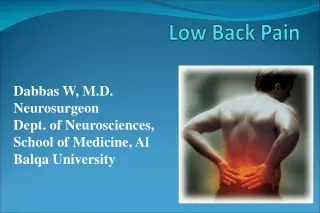
Clinically Effective Strategies for Common Presentations of Low Back Pain. Stephen Janz RN BN B Ac Cert Ac (China) GCCHM GCPH Fellow AACMA sjanz@kenmorehealth.com.au. Low Back Pain. Common and Debilitating

E N D
Clinically Effective Strategies for Common Presentations of Low Back Pain Stephen Janz RN BN B Ac Cert Ac (China) GCCHM GCPH Fellow AACMA sjanz@kenmorehealth.com.au
Low Back Pain • Common and Debilitating • Physiotherapy, Massage Therapy, Acupuncture, Point Injection Therapy, Exercise Therapy, Chiropractic, Osteopathy, Pharmacological Care. Surgery. • Inconsistent results from commonly used treatments in RCT’s • No Gold Standard
Programme • 10:00 Lecture: Pathology & Diagnosis • Practical: Soft tissue pelvic balance. • Lecture : Therapeutic approach to common presentations of low back pain. • 11:30 Break • 12:00 Demonstration of application of therapeutic methods. • Lecture: Rehabilitation. • 1:00 Close
Most Effective Treatment for Back Pain? • No more than 2 days bed rest • Keep active • 10 mg Morphine • Diazepam • Voltarin • ? acupuncture • Short term effect – pathology remains in every case. • Need a comprehensive problem solving approach
Pathology • Muscle strain • Muscle spasm • Facet Joint pathology • Sacro-illiac joint • Joint space narrowing and inflammation • Disc bulge • Disc Prolapse • Nerve/Nerve root compression • Trigger Points
Diagnosis • Acute episodes typically present without advanced diagnostic aids as such as Xray/CT/MRI. • Functional Diagnosis based on history, presentation , examination and response to treatment. • Specific cause not necessary to treat. • But need a working diagnosis for prognosis and patient confidence.
Pathology-Catagories • Facet Joint: 15% (40-50% in older populations) • Sacro-illiac Joint: 20% • Discogenic Pain: 40% • Non-Specific: 25% • Often mixed presentation
Presentation • Pain – • Local • referred • Radiating • Restricted function • Muscle Spasm • Inflammation
Presentation • Pain – local or radiating • Dull ,deep, burning , expanding, pressure like – more local: referred pain (somatic pain). • Sharp, shooting, electric; more distal than local – radicular pain – nerve root irritation. • Paraesthesia, numbness, tingling: -neuropathy – nerve root compression. • Positional aggravating and relieving factors • Other aggravating & relieving factors,
Presentation • Restricted function • Stiffness and discomfort on movement to severely compromised mobility • Muscle Spasm • Inflammation
Common Presentation • Lumbo-sacral pain +/- radiating to hip/groin/leg • Tenderness at level of L4/5 L5/S1 and Sacro-illiac joints • +/- local muscle Spasm • Pelvic Rotation (Psoas/Piriformis involvement) • Obvious altered Posture and guarded movement.
Sacroiliac Joint Pain • Pain below L5 – usually unilateral • Point to pain around hip/buttock • Tender Sacral Sulcus • Refers to thigh, lower leg, foot and ankle, groin and abdomen. • Often painful to stand from sitting “transition pain”
Facet Joint Pain • Commonly L4 and L5/S1 • Usually bilateral • Nonspecific tenderness infero-laterally from spinous process and over articular pillar
May be no back pain • Radiate to one or both legs. • Often worse with prolonged standing. • Easy to aggravate with manipulation. Discogenic Pain
Red Flags • Less than 1% sinister pathology • Sudden constant pain with no obvious cause • Failure to Improve • Pain unrelieved by rest at night • Refer to GP for investigations • ? tumours/infections/fractures.
Goal of treatment • Acute Phase • Reduce pain and inflammation • Restore structural balance • Stimulate repair • Restore function • Rehabilitative Phase • Identify and address underlying pathology • Identify & address predisposing lifestyle factors • Establish core stability programme • Prevent recurrence.
Acute Phase • Tools • Acupuncture (local +/- electro, auricular) • Analgesics • Pain impairs tissue repair and mobilisation (McCaffery & Pasero, 1999) • Anti-inflammatories • Antispasmodics • Soft-tissue techniques • Gain patients confidence with knowledge of condition & prognosis
Acute Phase • Assess for hip rotation and correct it • Anterior Superior Iliac Crest • Thumb length • Hip internal rotation supine. • Simple protocol often works • Often attacks the premorbid pathology • Reduces segmental strain and dysfunction. • Caution if suspect disc
Hip Balance • Start Supine • Lift backside to straighten spine • Compare ASIS, medial malleolus & thumb length • Release psoas on short side • Gentle ASIS rock • Quad traction on short side • Prone • Check hip internal rotation • Release with drainage/MTP/Bowen/Massage • If won't release suspect disc/ L2/L3 or hip degeneration
Acute Phase # 1 • Palpate lumbar and suitable massage to area (5-10 minutes) • If not much local spasm – choose local points egBl 25, 26, 27 and use electro stimulation at dense disperse about 4-100hz for 15 -20 minutes as long as is comfortable. • Look for trigger points in gluts egBl 54, Gb 29 • If waist involved – Bl40 • Palpate Bl 57 –use if tight/lump. • often Gb 34 bilaterally. • If Disc – Du mai SI 3 Bl 62. Scoliosis – Yang Wei mai Th5 Gb41
Acute Phase - 2 • If local spasm – no local points – auricular instead. – ear lumbar points selected by VAS. • Can procede with cautious massage to ease spasm but don’t over treat. • Distal pts as appropriate – often Gb 34 bilaterally. • If Disc – Du Mai SI 3 Bl 62 • Scoliosis -Yang Wei Mai TH5 Gb 41
Acute Phase - 3 • Don’t over treat. • The greater the pain and dysfunction – the less soft-tissue work and cautiously use local points (or avoid them). • Follow-up in 2 or 3 days. • Concurrent anti-inflammatory/analgesics if indicated for first few days.
Acute Phase 4 - advice • Warn the patient there may be transient aggravation • Advise them to take and anti=inflammatory or analgesic if you think you have over treated • Prescribe antispasmodic egUtramusclese/fibroplex/myoplex. • Give a clinical diagnosis • Reboook in 2 or 4 days
Anti-inflammatories/Analgesics& Antispasmodics • Reduce inflammation and pain • Promote mobilisation • Better sleep • Quicker recovery • Less socio-economic impact • Paracetamol, Ibruprofen, Voltarin, Panadeine etc. • Magnesium • Bromelains/Curcumin etc. • Contra-indications and side effects.
Auricular Acupuncture • Chinese and French are main systems • Since 1960’s. • Can be very intricate and uses the VAS pulse to identify best points. • Common locations often effective for low back pain.
Dry Needling=Trigger Point Acupuncture • Palpable nodule in a taught band • Twitch response to deep needling • Jump sign when needled (pt will call out – Ah Shi*!) • Deep or superficial needling – superficial safer and less painful. • Usual only 30 secs to 2 minute retention. • Can follow with stretch of treated area
Acupuncture Point Injection • Saline found to be as effective as other substances over 50 years ago. • Do not need to needle deeply (13 mm needle used) and therefore low risk. • Ideal for chronic/intractable and neuropathic pain. • ½-1 ml injected over jiaji/Bl or ah shi points. Choose segment above and below affected level.
Rehabilitative Phase • Identify and address underlying factors • Marked Degeneration on Xray – Glucosamine with chondroitin • Generalised stiffness – EPA (fish oil concentrate) • Lifestyle factors • Sedentary/seated/manual worker/sporting issues • Obesity • Core Stability
Core Stability • Transverse abdominis • Internal oblique • Multifidus • segmentally deactivated in back pain • Does not spontaneously activate • Learn yourself or refer to physio. • Exercise on the Ball • www.kenmorehealth.com.au/links core stability
Goal of treatment • Acute Phase • Reduce pain and inflammation • Restore structural balance • Stimulate repair • Restore function • Rehabilitative Phase • Identify and address underlying pathology • Identify & address predisposing lifestyle factors • Establish core stability programme • Prevent recurrence.
Clinically Effective Strategies for Common Presentations of Low Back Pain Stephen Janz RN BN B Ac Cert Ac (China) GCCHM GCPH Fellow AACMA Slides available at: www.kenmorehealth.com.au/links/seminar Password=janzseminar