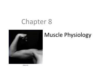
Muscle Physiology
Muscle Physiology. Lecture Outline. Muscle Function Muscle Characteristics Muscle Tissue Types Skeletal Muscle General Functions of Skeletal Muscle Functional Anatomy Physiology Skeletal Muscle Types Energetics Adaptive Responses Cardiac Muscle Physiology Smooth Muscle Physiology.

Muscle Physiology
E N D
Presentation Transcript
Lecture Outline • Muscle Function • Muscle Characteristics • Muscle Tissue Types • Skeletal Muscle • General Functions of Skeletal Muscle • Functional Anatomy • Physiology • Skeletal Muscle Types • Energetics • Adaptive Responses • Cardiac Muscle Physiology • Smooth Muscle Physiology
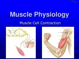
Muscle Function • Movement • Depends on type of muscle tissue • Depends on location of muscle tissue • Thermogenesis • Protection • Posture Maintenance • Joint Stabilization
Muscle Tissue Characteristics All muscle tissues share basic characteristics • Excitability • Contractility • Elasticity • Extensibility
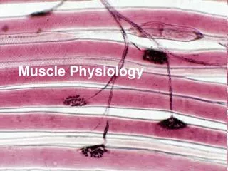
Muscle Tissue Types Skeletal Cardiac Smooth
Muscle Comparison Chart Muscle Tissue Special structures Striae Cell Shape Nucleus Control Multi-nucleate & peripheral Skeletal Cylindrical Yes Voluntary none Intercalated discs Uninucleate & central Cylindrical & branched Cardiac Involuntary Yes May be single-unit or multi-unit Uninucleate & central Involuntary No Fusiform Smooth
Skeletal MuscleGeneral Functions - Voluntary • Movement • Only have contractility in one direction • Requires multiple muscles to create movements from the simple • flexion and extension • To the complex • Circumduction • Stabilizing Movements & Joints • The result of synergistic muscles
Skeletal MuscleGeneral Functions • Protection • of underlying structures • abdominal viscera • Stronger muscles = greater protection, increased joint stability
Skeletal MuscleGeneral Functions - Involuntary • Shivering Thermogenesis (shivering reflex) • asynchronous & involuntary • Initiated by hypothalamic nuclei in the primary motor center for shivering (posterior nuclei) • Normally inhibited by the heat center in the hypothalamus (preoptic nuclei)when body temp is in range (96.8-99.5) • Receives cold signals from skin and spinal cord Skeletal Muscle - - posteriornucleus preoptic nucleus Damage to the posterior nuclei would cause?
Skeletal MuscleGeneral Functions - Involuntary • Maintenance of Posture • Involves stretch reflexes • Static reflexes • Long term sustained contractile events • Phasic reflexes • Dynamic and short term corrective responses • Regulated by gamma neurons which adjust tension in the muscle spindles
Skeletal MuscleFunctional Anatomy • The smallest functional unit of skeletal muscle is the sarcomere
Skeletal MuscleFunctional Anatomy • Sarcomere is composed of various microfilaments and supporting structures • Titin • largest known elastomeric protein • Connects myosin to z-disc • thought to be critical in the development of sarcomeres
Skeletal MuscleFunctional Anatomy • Myosin molecule consists of tail, hinge and heads • Heads contain active sites for • Actin • ATP • M-line consists of myomesin and skelemin proteins • stabilize the myosin filaments • theorized to aid in transmission of force from sarcomere to cytoskeletal intermediate filaments
Skeletal MuscleFunctional Anatomy • Thin filaments are composed of • g-actin molecules in a helical arrangement • Contain myosin binding sites • nebulin • Filament that formsinternal support andattachment for actin • tropomyosin filaments • troponin (complex of three molecules)attached to tropomyosin • Has binding sites for Ca2+
Skeletal MuscleFunctional Anatomy • The Z-disc • Anchors the filaments and interacts with cytoskeletal framework
Skeletal MuscleFunctional Anatomy • Transmission of force from the sarcomere to the tissue at large • Sarcomeres linked by dystrophin to sarcolemma, then via membrane proteins interacting with cytoskeletal framework Muscular Dystrophy?
Skeletal MusclePhysiology of Contraction • How does all this functional anatomy work? • 1st – synaptic transmission at the neuromuscular junction • 2nd – excitation-contraction coupling • 3rd – contraction-relaxation cycle
Skeletal MusclePhysiology of Contraction - NMJ • Events at the neuromuscular junction (NMJ) • action potential arrives at the pre-synaptic membrane • depolarization of membrane opens voltage gated Ca2+ channels • calcium influxes into synaptic bulb • calmodulin is activated by Ca2+ which • activates protein kinase II (PK II) • PKII phosphorylates synapsin (motor protein) • vessicle binds to membrane proteins (SNAREs) • exocytosis of ACh • ACh binds to nicotinic receptors • Na+ influx creates an End Plate Potential (EPP) • EPP spreads to edge of the motor end plate and initiates an action potential in the sarcolemma
Skeletal MusclePhysiology of Contraction – Excitation-Contraction Coupling • Excitation-Contraction Coupling Process • Action potential spreads along sarcolemma and down t-tubules • Depolarization of membrane alters membrane protein dihydropyridine L (DHP) configuration • Altered DHP configuration signals ryanodine Ca2+ receptors (RyR Ca2+) in the terminal cisternae of the sarcoplasmic reticulum • Neatly, these are near the I and A bands of the sarcomere! • Ca2+ is released into the sarcoplasm and • binds to troponin • initiates a conformational change in the troponin-tropomyosin complex exposing the binding sites for myosin on actin • Myosin binds to actin (electrostatic attraction)
Skeletal MusclePhysiology of Contraction – Contraction-Relaxation Cycle • Contraction-Relaxation Cycle • Myosin upon attaching to actin is hydrolized (phosphate coming from the splitting of ATP by Myosin ATPase) • This changes the conformation of myosin causing it to bend at the neck towards the m-line • ADP is released by the conformational change during the “power stroke” • ATP binding site is now available for another ATP (along with magnesium Mg2+) • Splitting of ATP to ADP + P by myosin detaches and returns myosin to its active state • This single event creates a twitch
Skeletal MusclePhysiology of Contraction • Animation of Skeletal Muscle Contraction-Relaxation Events
Next Time… • Muscle Energetics • Muscle types