Download

1 / 35
350 likes | 567 Views
William 2001. Nonimmune hydrops hemorrhagic diseases of the newborn. Hyperbilirubinemia Nonimmune hydrops Cardiac arrhythmias Hemorrhagic disease of the newborn Thrombocytopenia Polycythemia Necrotizing entrocolitis. Unconjugated bilirubin : Not excreted in bile and urine

E N D
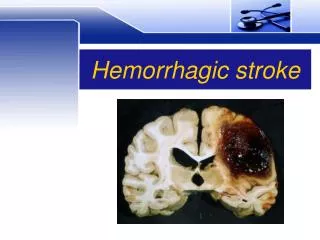
William 2001 Nonimmunehydropshemorrhagic diseases of the newborn
Hyperbilirubinemia • Nonimmunehydrops • Cardiac arrhythmias • Hemorrhagic disease of the newborn • Thrombocytopenia • Polycythemia • Necrotizing entrocolitis
Unconjugatedbilirubin: • Not excreted in bile and urine • Pass the placenta to the mother Conjugated bilirubin: • Water soluble • Excreted in bile and urine Kernicterus: ↑ unconjugatedbilirubin > 18 – 20 mg/dL - < 18 in preterm hyperbilirubinemia
Clinical picture: • Spasticity • MR • Muscle incoordination Causes of kernicterus: • Hypoxia • Hypoglycemia • hypothermia • Acidosis • sepsis
Drugs: Furosemide Gentamicin Salicylates Sulfonamides Diazepam Na benzoate ↑ vitamin K1
Brest milk jaundice: - Due to excretion of: pregnane – 3 α, 20 β–diol in the milk inhibit conjugation of bilirubin by inhibiting glucuronyltransferase activity - Jaundice starts 4th to 15th day - No encephalopathy
Physiological jaundice: Starts 3rd to 4th day Bilirubin level < 10 mg/dL Phototherapy: For treatment of hyperbilirubinemia Mechanism: Ligh oxidation of bilirubin ↓ ↑ peripheral blood flow ↑ photooxidation
Method: • Eyes covered • Skin exposed • Appropriate fluorescent wavelength • Baby turned /2 hours • Bilirubin measured after 24 hours • Monitor temperature to prevent dehydration
Definition: Abnormal fluid accumulation in ≥ sites Incidence: 0.6 % 77% of them are known 1.7 % 95% of them are known Incidence of hydrops: 13% immune 1.3% extrinsic 21% idiopathic 64% intrinsic Nonimmunehydropsfetalis
Intrinsic causes: 41% cystic hygroma 27% cardiac anomalies 21% multiple malformations 11% others Causes of nonimmunehydrops: 1 – Cardiac: = 20 – 45%
½ structural anomalies ½ cardiac arrhythmia 2 – Chromosomal anomalies: = 35% - earlier - extensive space suite hydrops 87% with anencephaly 3 – Severe anemia: Parvovirus Acute fetal - maternal Hg α - thalassemia
4 – Twin-to-twin transfusion: Recipient HF Donor hydrops after the death of the recipient 5 - Inborn errors of metabolism: - Gaucher disease - GM 1 gangliosidosis - Sialidosis All recurrent hydrops
6 – Lymph system anomalies: - Chylothorax - Chylousascites Prognosis: < 24 weeks 95% mortality ≥ 24 weeks 80% mortality Diagnosis: Maternal tests – cordocentesis - US
Maternal tests: • Hb electrophoresis • Indirect Coombs test • Kleihauer – Batke test • Serological tests for: Rubella Toxoplasmosis Syphilis Cytomegalovirus Parvovirus B - 19
Cordocentesis: • karyotyping • Hb% • Hb electrophoresis • Direct Coombs test • Liver transaminases • Serological test for Ig M specific Abs
Most important predictor tests for prognosis: • Karyotyping • Fetal ECG Management: - Blood transfusion for anemia - Amniocentesis for twin-to-twin transfusion may spontaneous cure If persistent exclude cardiac anomalies and anencephaly
Deliver if near term Expectant treatment if very preterm Maternal complications: • Mirror syndrome: Edema and preeclampsia due to vascular changes in the fetus • Others: Overdistension PTL – PP Hg -- retained placenta
Usually transient and benign Some tacchycardia if sustained may hydrops, HF and fetal death Sustained bradicardia is caused by: • Congenital anomalies • Myocarditis And is less often associated with hydrops Cardiac arrhythmias
Isolated extrasystoles: • Atrialextrasystoles • Ventricular extrasystoles Sustained arrhythmias: • Supraventriculartacchycardia • Ventricular tacchycardia • Complete heart block • 2 degree heart block • Atrial flatter, fibrillation • Sinus bradicardia Types of arrhythmias
Premature atrial contractions: = 64% of cardiac arrhythmia Usually benign and transient Rarely supraventricular tacchycardia and if > 200 b/m may HF Bradicardia: Poor prognosis Caused by:
Structure anomalies as A-V canal • Heart block Congenital heart block: - Caused by Abs against fetal myometrium in 50% of the cases - Most common Abs: Anti-SS-A (Anti Ro) Abs - Inflammation and permanent damage to the myocardial tissue
- Neonate may require pacemaker - Only 1 : 20 of the cases are affected - Mothers usually have: SLE or other CT disease or subsequently develop it Fetotherapy : By corticosteroids to the mother
Characterized by: • Hypoprothrombinemia • ↓ factor V, VII, IX, X • ↑ prothrombin time • ↑ PTT Spontaneous internal or ext Hgs May occur at any time Usually delayed 1 – 2 days Hemorrhagic disease of the neonate
Causes: • ↓ vit k1 • Hemophilia • Sepsis • Syphilis • Thrombocytopenia • Erythroblastosis • ICH
Vit K1: • ↓ during pregnancy # nonpregnant • ↓ placental transmission • ↓ in milk Anticonvulsive drugs prevent hepatic synthesis of factor VII, IX, X ↓ vit K1 A phenotype similar to Chondrodysplasia punctata = Conradi – Hunermann syndrome =inherited disease characterized by bone dystrophy and facial anomalies
Types: 1 – Immune thrombocytopenia: - Maternal antiplateletIg G fetal/ neonatal thrombocytopenia - Usually associated with maternal autoimmune disease and maternal thrombocytopenia - Corticosteroid therapy ↑ maternal platelet count but does not improve fetal condition thrombocytopenia
2 -- Alloimmune thrombocytopenia (ATP): - Fetal platelet Ag pass the placenta to the mother isoimmunization - Usually discovered after the delivery of an affected child - May IC Hg - 98% of the population are HPA 1a +ve 2 % of the population are HPA 1a –ve - % = 1 : 5000 – 10000 live birth - 1 : 50 of pregnancies are at risk
- Significant fetal – maternalHg must occur provoke immune respond - Affect offspring of women with HLA type DR - 3 or B - 8 Diagnosis: - Maternal platelet count normal +no autoimmune disease - Fetal platelets count ↓ + no other autoimmune D
- IV injection of Ig in a large dose to the mother recurrent fetal thrombocytopenia by cordocentesis Recurrence = 70 – 90% More severe and earlier in subsequent pregnancies
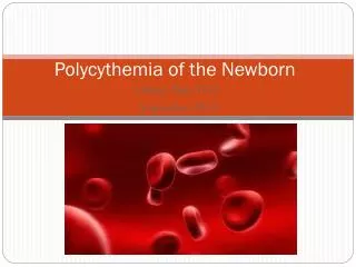
Predisposing factors: • Chronic hypoxia • Placentaltransfusion ( maternal or twin) Clinical picture: • Plethora • Cyanosis • Neurological impairment polycythemia
Laboratory: • ↑ bilirubin • ↓ platelet • Hypoglycemia • Fragmented RBCs Treatment: plasma
Bowel disorder affects mainly premature neonates due to intestinal immaturity Clinical picture: • Distension • Illus • Bloody stools X ray: Gas in intestine = pneumatosisintestinalis May perforation Necrotizing enterocolitis
% 5.7of preterm infants Causes: • Perinatal hypotension • hypoxia • Sepsis • Umbilical catheters • Exchange transfusion • Hypertonic fluids
Cow milk • Coronovirusinfection Treatment: Ig administration orally