Download
1 / 20
200 likes | 324 Views
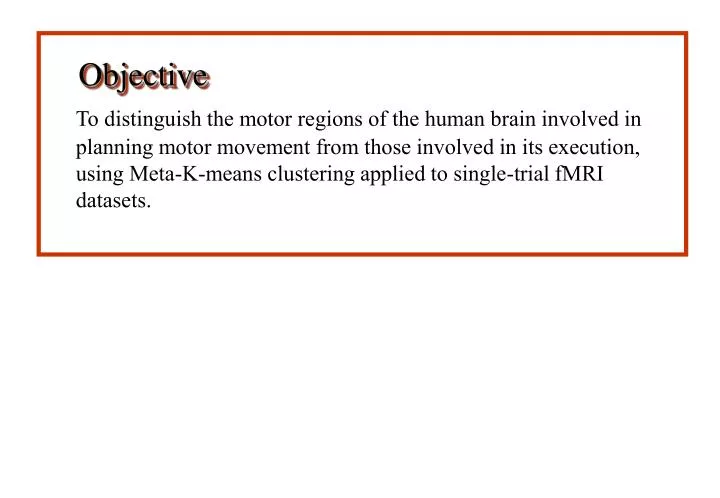
Objective. To distinguish the motor regions of the human brain involved in planning motor movement from those involved in its execution, using Meta-K-means clustering applied to single-trial fMRI datasets. Data Acquisition: Materials and Methods.

E N D
Objective To distinguish the motor regions of the human brain involved in planning motor movement from those involved in its execution, using Meta-K-means clustering applied to single-trial fMRI datasets.
Data Acquisition: Materials and Methods • Single-shot EPI images using a 4 Tesla MRI system • Normal adult, right-handed human subjects • 10 coronal sections of thickness 5 mm each, including the primary, supplementary and pre-motor areas • 64 x 64 images, acquired at the rate of one volume per second
Paradigm A delayed, cued joystick movement task: The subject was asked to move a cursor controlled by a joystick, from the centre of the screen to the memorized location of a target. (Fig. 1)
Cursor moves out of centre: Beginning of execution phase Target appears: Beginning of preparation phase Fig. 1
The cursor was positioned in the centre of the screen at the beginning of the task. A yellow target appeared very briefly at one of 8 possible locations close to the periphery of the screen. Disappearance of the target was followed by a pseudo-randomized variable (0-3 seconds) delay period, the end of which was signalled by the change in colour of a circle at the centre, from red to green (the ‘go’ cue). The subject was to then move radially to the memorized location of the target, as accurately and quickly as possible. Response accuracy, reaction and movement times were recorded for each trial.
One continuous imaging sequence (run) consisted of 16 single trials, with intertrial periods of 20 seconds each. Between the end of one execution phase and the beginning of the next trial (i.e. the control, or no-movement period), the same visual stimulus (flashing targets) was presented, but the ‘go’ cue was not given. A total of 340 volumes were acquired in each run.
Data Analysis Preprocessing Lyngby fMRI Analysis Toolbox Initial K-means clustering Select 2-3 clusters FIR filtering Impulse response function Meta-K-means clustering
Initial K-Means Clustering: Each slice was processed separately. The voxels in a slice were represented as vectors in 320-dimensional (no. of time-points) space. Each such vector was normalized to unit length, and K-means clustering performed. The resulting clusters were examined visually. Clusters which showed a relatively clean time-course, as well as were reasonably spatially confluent, were chosen for further analysis. This typically was 2-3 clusters per slice. Preprocessing: • Discarding the first 20 pre-steady-state volumes • Zeroing the mean of each trial in a run
Finite Impulse Response Filtering: The voxels belonging to the chosen clusters were each deconvolved with two different paradigm functions to get the respective finite impulse responses (FIR’s) of each. Meta-K-Means Clustering: The FIR filter waveforms were normalized to unit vectors after DC shifting each to make the coefficients non-negative. K-means clustering was again performed on these filters. • The 2 paradigm functions were : • Preparation: target ‘on’ to beginning of movement • Execution: beginning to end of movement
Initial K-Means Clustering: Choosing the value of K: The intracluster variance averaged over all pixels, was used as the cost function. In general, this is a decreasing function of the number of clusters, with an increasing slope. (Fig. 2) A dip in the slope of this function, indicating a more rapid decrease of the variance for that particular cluster configuration, was used to choose the value of K. Results
Variation of intracluster variance (averaged over all voxels) as number of clusters is increased : 6, 7 and 10 were chosen as possible values of K in this case. Fig. 2
R L Selection of clusters for further analysis: Fig. 3 shows two clusters from a single coronal slice, that were selected for further analysis based on their mean time courses and spatial distribution of voxels.
Two clusters of voxels that resulted from initial K-means clustering; the mean time-course of each of these clusters is shown alongside Fig. 3
FIR Filtering Picking the length of the FIR filter: The FIR filter was chosen to be long enough to allow the waveform to return to baseline in representative voxels in the motor cortex. (Fig. 4) The FIR filter waveform can be considered to represent the BOLD response of the particular voxel. Thus, haemodynamic activity appears to last for ~18 seconds in the motor areas following the beginning of a trial.
FIR filters of different sizes, for a representative voxel in the motor cortex: a filter of length 18 was chosen to cover the entire BOLD waveform in different voxels Fig. 4
Meta-K-Means Clustering Examples of clusters that resulted from meta-K-means clustering are shown in Figs. 5(a) and (b). The cluster in Fig. 5(a) includes voxels from bilateral primary and pre-motor areas, and the cerebellum. The preparation paradigm was used here. The mean FIR filter shows activation immediately after target presentation, i.e. in the preparation period. The first preparation peak (arrow) is followed by a second, larger peak very closely, which is likely due to motor execution. The cluster in Fig. 5(b), on the other hand, resulted from filtering with the execution paradigm, and consists mainly of draining veins. The mean filter shows a late rise and a broad BOLD waveform, as would be expected in a vein.
R L 5(a): A meta-K-means cluster obtained by filtering with the preparation paradigm. The mean FIR filter of this cluster shows an early preparatory activation (arrow) that is partly masked by the larger execution-related activation.
5(b): Another meta-K-means cluster, obtained by filtering with the execution paradigm. The mean FIR filter here shows a delayed and broad response that likely represents the signal from draining veins. Fig. 5
Conclusions: The voxels in the motor areas clearly showed differences in activation in terms of temporal relation to the planning and execution phases of a motor task. FIR filtering followed by K-means clustering on the coefficients provides a good tool not only to separate voxels of brain tissue with different temporal activation patterns, and veins in single-trial experiments, but also to visualize the BOLD response waveform.
References: • Goutte, C., Toft, P., Rostrup, E., Nielsen, F. Å., and Hansen, L. K. 1999. On Clustering fMRI Time Series. NeuroImage9, 298-310 • Nielsen, F. Å., Hansen, L. K., Toft, P., Goutte, C., Mørch, N., Svarer, C., Savoy, R., Rosen, B., Rostrup, E., Born, P. 1997. Comparison of Two Convolution Models for fMRI Time Series. Third International Conference on Functional Mapping of the Human Brain. (Friberg et. al., Eds.) NeuroImage3(3), S473 • Ripley, B. D. 1996. Pattern Recognition and Neural Networks. Cambridge University Press Supported by NIH (MH57180 and RR08079)