Download
1 / 14
180 likes | 581 Views
Canine Paraprostatic Cyst. Accession #’s: 80443, 80442, and 80468 Christina Copple, DVM April 27, 2009 Radiology Resident NCSU CVM-VTH. Jake 9yr old M German Shepherd. rDVM in June 2007 straining to defecate Prostatomegaly and prostatic cyst were diagnosed

E N D
Canine Paraprostatic Cyst Accession #’s: 80443, 80442, and 80468 Christina Copple, DVM April 27, 2009 Radiology Resident NCSU CVM-VTH
Jake 9yr old M German Shepherd • rDVM in June 2007 straining to defecate • Prostatomegaly and prostatic cyst were diagnosed • Seen by NCSU CVM-VTH surgery service July 2007 • Still straining to defecate but no difficulty urinating • 6 lb weight loss and diarrhea since June • Enlarged and painful prostate on rectal • Azotemia • HX of arthritis
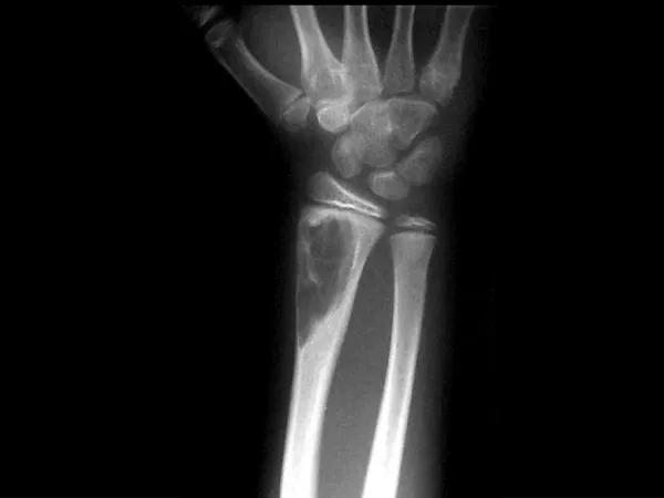
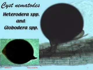
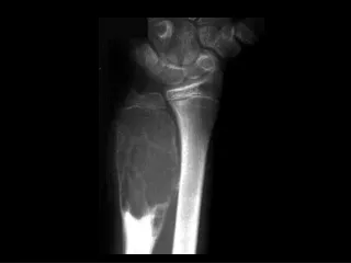
Soft tissue mass caudodorsal to urinary bladder Accession #80443
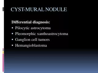
Large paraprostatic cyst from right lobe of prostate gland Large, heterogeneous prostate gland Accession #80442
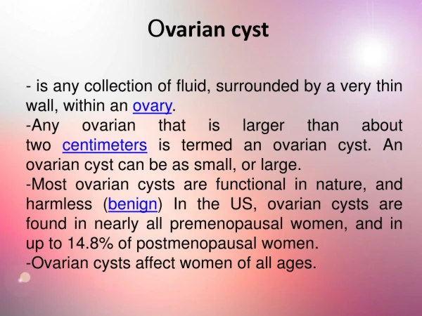
Positive Contrast • Cystourethrogram: • No evidence of communications • between paraprostatic cyst and urethra • Mild prostatic reflux of contrast medium Accession #80468
Canine Paraprostatic Cyst(Renfrew, Barrett, Bradley, and Barr; VetRadUS 49:5, 2008. 444-448) • Relatively uncommon and cause often difficult to determine • Ductal occlusion secondary to squamous metaplasia → progressive secretory stasis (multiple cavitary areas within gland or single large fluid-filled structure in abdomen) • End-stage prostatic hematoma with mineralization of fibrous wall • Originate from uterus masculinus
Canine Paraprostatic Cyst(Renfrew, Barrett, Bradley, and Barr; VetRadUS 49:5, 2008. 444-448) • Mineralization is uncommon but can occur • Differentiate between prostatic retention cysts vs paraprostatic cysts? • Prostatic retention cyst = intimate association with prostate gland, often communicates with the urethra • Paraprostatic cysts = only minimal structural communication with prostate gland, no urethral communication http://www.best-dog-photos.com/images/German-Shepherd-Dog.jpg
Radiographic and Ultrasonographic Features of Canine Paraprostatic CystsRenfrew, Barrett, Bradley, and Barr; VetRadUS 49:5, 2008. 444-448 • Retrospective, 8 dogs, 3-8 yrs of age, all medium to large breed • Boxers (5/8 = 62.5%), 1 each of GSD, German Shorthaired Pointer, and Irish Wolfhound • Clinical signs: • 7 – related to urinary tract and/or defecation (1 – systemic illness, 1 – hindlimb lameness) • 1 – none
Radiographic and Ultrasonographic Features of Canine Paraprostatic CystsRenfrew, Barrett, Bradley, and Barr; VetRadUS 49:5, 2008. 444-448 • Prostatomegaly = craniocaudal dimension of the gland being > 70% of the distance between the sacral promontory to the pubic brim on a lateral radiograph • Radiographically: • All had dorsal displacement of the colon and/or rectum • Displacement of urinary bladder, small intestine • ½ had radiographic evidence of cyst mineralization; pattern of mineralization was varied
Radiographic and Ultrasonographic Features of Canine Paraprostatic CystsRenfrew, Barrett, Bradley, and Barr; VetRadUS 49:5, 2008. 444-448 • Ultrasound was performed in 7 of 8 dogs • All had hyperechoic rims and hypo- or anechoic contents • Other findings: septations, stalk, mineralization • Communication or stalks between the paraprostatic cyst and the prostate gland were identified in 7 of 8 dogs • All 8 dogs had histopathology consistent with paraprostatic cyst
Radiographic and Ultrasonographic Features of Canine Paraprostatic CystsRenfrew, Barrett, Bradley, and Barr; VetRadUS 49:5, 2008. 444-448 Conclusions: • Cyst location was determined by a combination of imaging & surgery • When prostatic cyst was not mineralized contrast radiography was necessary to allow differentiation of cyst from bladder; provides a global view and allows for determination of communication of prostatic cyst with urethra • Ultrasonography is a valuable tool but can be limited if used alone; may not document mineralization if focus of mineralization is <5 mm tall • For complete characterization of prostatic cyst recommend retrograde urethrography and ultrasound • Paraprostatic cyst are frequently mineralized with superior detection via radiography when compared to ultrasound
What about Jake? • Surgery: scrotal ablation, left inguinal herniorrhaphy, resection of paraprostatic cyst and prostatic biopsy • BX = benign cystic prostatic hyperplasia with mild lymphoplasmacytic inflammation • Five days post discharge Jake was doing well at home, recommended recheck ultrasound in 3 months, lost to follow-up http://germanshepherddog.ca/images/funnyface.jpg
Reference • Renfrew, Helen, Esther L. Barrett, Kate J. Bradley, and Frances J. Barr. Radiographic And Ultrasonographic Features Of Canine Paraprostatic Cysts. VetRadUS, 49:5, 2008. 444-448