Unraveling the Mystery of Unique Phage Growths: A Personal Study
This report by Ashley Bue details the intriguing findings from the analysis of phage samples collected from her hometown in Minnesota. With a focus on bacterial growths observed on plaques and the development of a biodegradable film covered with phages, this study highlights significant implications for medical applications, particularly in wound care and burn treatment. The work underscores the importance of phages in our environment and reveals the personal journey of exploration in the field of microbiology, driven by the influence of a dedicated high school biology teacher.

Unraveling the Mystery of Unique Phage Growths: A Personal Study
E N D
Presentation Transcript
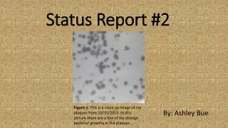
Status Report #2 By: Ashley Bue Figure 1: This is a close up image of my plaques from 10/15/2013. In this picture there are a few of my strange bacterial growths in the plaques.
Phage Fun Facts • Phages’ play a essential role in the environment and our human lives. • Currently, there are a few selectively studies working to develop current medical conditions. • One of these studies has developed a biodegradable film that is covered with phage. This film has been successful used to cover wounds and prevent bacterial infections. • Also, there is treats using phages being developed for burn victims. Reference: Kirby, Breeann, and Jeremy J. Barr. "Going Viral." The Scientist. www.the-scientist.com, 1 Sept. 2013. Web. 04 Dec. 2013.
Phage Name: Gathje • Named after my High School Biology Teacher, Rochelle Gathje. • She influenced me to pursue a career in biology and through her classes I found my passion for biology. • She even graduated from UWRF! Figure 2: University of Wisconsin-River Falls mascot.
Source of Sample • On 9/8/13 I collected my sample from under a pinetree in a flower bed with cedar mulch. The top soil appeared dry and it contained numerous roots and pieces of mulch. • The weather was about 70⁰ F with an overcast sky but not raining. • GPS Coordinates: 43⁰ 49’ 31 N 91⁰ 58’ 21” W • 10 Minutes outside of Lanesboro, MN Figure 3: Pictured is my hometown of Lanesboro, MN
Development of Plaques Figure 7: 10/15/13 plaques which contain no bacterial growth. Figure 4: These are my first major plaque growths with the bacterial spots. Plaques from 9-26-13 Figure 5: Plaque from 10/3/12 that contains a bacterial growth. Figure 6: Spot test from 10/8/13 all plaques contain the bacterial growth. Figure 8: Plaques from 10/12/13 show growth again after redoing previous procedure because of contamination. Figure 9: Plaques from 10/22/13, one of my final plates.
Plaque Morphology • Titer of HTL: 5.4x10^9 • Size: 8 to 10 mm • Appearance: Clear, Round • Had Strange Appearance to Start Figure 9: This is my 10^ -5 HTL Titration plate created on 10/22/2013.
DNA Isolation • Concentration: 0.1775 μg/μl • Yield: 17.75 μg • A260/280: 1.699 • The ratio of A260/280 should be around 1.8 so mine is a little low but the number is still good. • I have enough DNA to preform all procedures.
Restriction Enzyme Results #1 Gel Electrophoresis #1 1 Kb Ladder Undigested DNA BamHI ClaI EcoRI HaeIII HindIII Figure 10: My first gel results from 10/31/13.
Analysis Fragment Lengths for Restriction Enzymes #1 • When looking at this gel, I can determine that my phage is possible related to three other phages in the class, SJD, JD, and TT. • With only these results it is hard to determine which ones are related or the same phage. • By studying the phages further I should be able to determine which ones are related. Figure 11: This chart depicts my fragment length from my first gel.
Restriction Enzyme Results #2 Gel Electrophoresis #2 PST I SAL I ECO RV NCO I KB Ladder Figure 12: This is my second gel from 11/12/13.
Fragment Length for Restriction Enzymes #2 Figure 13:
Analysis of Second Set of Restriction Enzymes • From the second gel, I am able to conclude that it is not related to SJD and JD. • My gel is completely different from both of these gels, both are picture below. Figure 14: JD gel had different cuts than mine and SJD. Figure 15: SJD gel had different cuts than mine and JD.
Electron Microscope Results • 1, 2 are normal siphoviridae phages. • 3 is missing the DNA inside its head. • 4 has a broken open head which has allowed the dye to enter. • 5 is a normal siphoviridae phage with a curled tail. 2 Phage 1: Tail-96.88nm Head-43.75 nm Phage 2:Tail-100nm Head-40.63nm Phage 3:Tail-100 nm Head-43.75 nm 1 4 5 3 Figure 16: This is my image from the electron microscope.
Final Conclusions • For all of this information I can conclude that my phage is for sure not related to JD or SJD because of the second gel. • A third gel was ran to determine if mine and TT were related. This gel showed very poor results so I can not determine if the two are related. Also there is not a good electron microscope image of TT so I can’t clearly determine if the two images look similar. • My phage shows no exact similarities to any sequenced phage in the database.
Interesting! • My phage started to develop strange bacterial growths in the center of my plaques. • Slowly these growths started to disappear as a purified the phage. Figure 17: This is a streak plate created from the bacterial growths in the center of my plaqes.
WHY MY PHAGE! • Unique bacteria growth! • I have enough DNA. • I have take accurate notes on all procedures. • My phages is not taken from on of Karen’s samples.