Download
1 / 26
290 likes | 657 Views
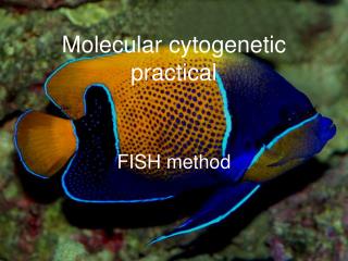
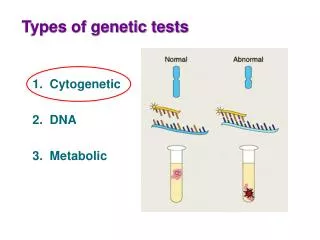
Types of genetic tests. 1. Cytogenetic 2. DNA 3. Metabolic. Karyotype Picture of the chromosomes in a cell used to check for abnormalities. Prenatal diagnosis: trisomy 21 (Down’s syndrome). Postnatal diagnosis:. Postnatal diagnosis: detecting cancer. Preparing a karyotype

E N D
Types of genetic tests 1. Cytogenetic 2. DNA 3. Metabolic
Karyotype • Picture of the chromosomes in a cell used to check for abnormalities Prenatal diagnosis: trisomy 21 (Down’s syndrome)
Preparing a karyotype • Harvest cells (from where?) Postnatal diagnostic karyotype Prenatal diagnostic karyotype
Preparing a karyotype • Harvest cells Postnatal diagnostic karyotype • tumor biopsy • skin cells from mouth (ie for non-cancer related diagnoses) Prenatal diagnostic karyotype • chorionic villi sampling (CVS) • amniocentesis
Who is offered amniocentesis or CVS? • Maternal age (women 35 or older) Risk of Down’s syndrome: mother in 20s 1/1250 99.92% OK mother at 35 1/400 99.75% OK mother at 40 1/100 99% OK
Down’s syndrome: how does it happen? Chromosomal non-disjunction during meiosis of eggs and sperm. 1. Chromosomes replicate 2. Homologous chromosomes separate 3. Chromatids from each chromosome separate
Who is offered amniocentesis or CVS? • Maternal age (women 35 or older) • Risk of Down’s syndrome: mother in 20s 1/1250 99.92% OK mother at 35 1/400 99.75% OK mother at 40 1/100 99% OK • A previous child or pregnancy with a birth defect • Screening test with a positive result • Other family history
Prenatal diagnosis: amniocentesis • Sampling cells from amniotic fluid • Usually done ~ 15–18 weeks
Prenatal diagnosis: chorionic villi sampling (CVS) • Sampling cells from placenta • Usually done 10–12 weeks
Preparing a karyotype • Harvest cells • Culture cells 1–2 days • Arrest cells in metaphase withcolchicine metaphase
Mitosis chromosomes condense DNA replication nuclear envelope breaks down metaphase chromosomes aligned on spindle fibres
Preparing a karyotype • Harvest cells • Culture cells 1–2 days • Arrest cells in metaphase with colchicine • ‘Spread’ cells on slide and stain • Count chromosomes in 20 representative cells • Capture image of five ‘best’ cells and construct karyotypes for each metaphase
FISH analysis of chromosomes: Fluorescent In Situ Hybridization Metaphase spread chromosomes stained with DAPI, a fluorescing stain that specifically binds double-stranded DNA
FISH Expose DAPI-stained metaphase chromosomes to fluorescent probes red = control probe for centromere of the X chromosome and another probe for end of chromosome X green = probe for the end of chromosome 4
DiGeorge syndrome/CATCH22 • Microdeletion on chromosome 22 • Birth defect that affects the immune system • Absence or underdevelopment of the thymus and parathyroid glands • Facial features include low-set ears, wide-set eyes, small jaw and bowing up of upper lip
FISH tests: DiGeorge syndrome Expose DAPI-stained chromosomes to mixture of fluorescent probes green = control probe for chromosome 22 red = probe for DiGeorge region on long arm of chromosome 22
FISH tests: Painting chromosomes Expose chromosomes to fluorescent probes that highlight entire chromosomes.
FISH tests: Painting chromosomes Expose chromosomes to fluorescent probes that highlight chromosomes 13, 18, 21, X and Y. nuclei from the same foetus green = chromosome 13 red = chromosome 21 aqua = chromosome 18 green = X chromosome red= Y chromosome
Trait Aphysical characteristic that is determined by genes, eg eye colour.
Human traits Thumb shape hh Hh or HH ‘Hitchhiker’s thumb’ Earlobe attachment AA or Aa aa unattached attached
Human traits Thumb shape hh Hh or HH ‘Hitchhiker’s thumb’ Genotype vs phenotype Genotype = specific allelic make-up of an individual, eg HH, Hh or hh Phenotype = an observable physical or measurable biochemical characteristic, eg thumb shape or lack of a particular enzyme
Punnett squares Remember these?? Used to determine the probability of an offspring having a particular genotype hh Hh or HH H H ‘Hitchhiker’s thumb’ H h H allele = dominant h allele = recessive
Punnett squares Now try it backwards hh Hh or HH ‘Hitchhiker’s thumb’ Hh Hh H allele = dominant h allele = recessive hh hh
Recombination: Shuffling the deck DNA crossovers in chromosome pairs that result in children receiving a different combination of genes than either parent