Download

1 / 18
180 likes | 323 Views
Physiological bases of hemo dynamic. Kinds of blood movements. Formulas of hemodynamic. Functional types of vessels. Amortization or compensatory vessels Resistive vessels. Functional types of vessels. Sphincters Capillars. Functional types of vessels. Volume vessels Shunts.

E N D
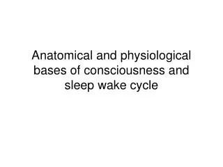
Functional types of vessels • Amortization or compensatory vessels • Resistive vessels
Functional types of vessels • Sphincters • Capillars
Functional types of vessels • Volume vessels • Shunts
Arterial pressure • Determine the influences of factors: • 1. cardiac – systolic volume, speed of blood ejection from the ventricles, heart beat; 2. vascular–elastisity of compensatory arteries, tone of resistive vessels, volume of volume vessels; • 3. blood–volume of blood, viscosity, hidrostatic pressure of blood.
Kinds of arterial pressure • 1. Systolicor maximal • 2. Side or absolute systolic • 3. Stroke (hemodynamic) • 4. Diastolic or minimal • 5. Pulse • 6. Result– • де Р –middledynamic pressure; Pd –diastolic pressure; Pc –systolic pressure. • Ideal prwessure: • Systolic = 102 + (0,6 · age) mm Hg • Diastolic = 63 + (0,4 · age) mm Hg
Sphygmogram Anacrota -а Catacrotab Incisura (i) Addition wave с or secondary increase
Arterial pulse • А.radialis • A. ulnaris • A. brachialis • A. carotica communis • А. temporalis • A. femoralis • A. dorsalis pedis • A. tibialis posterior 5 4 3 1 2 6 8 7
Basal tone of vessels. • When arterial pressure suddenly increases local blood flow tends to increase. Than local blood flow decreases to normal level. Vessel walls are capable to prolonged tonic contraction without tiredness even at rest. Such a condition is supported by spontaneous myogenic activity of smooth muscles and efferent impulsation from autonomic nerve centers, which control arterial pressure. Partial state of contraction in blood vessels caused by continual slow firing of vasoconstrictor area is called vasculomotor tone.