Download
1 / 39
400 likes | 668 Views
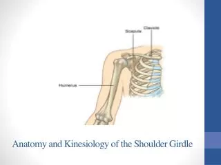
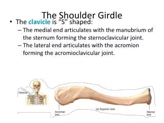
Shoulder Girdle. Tanya Nolan. Shoulder Girdle. Formed by 2 bones Scapula Clavicle Function Connect upper limb to trunk. Clavicle. Long Bone Horizontal oblique plane Doubly curved for strength Function Fulcrum for movements of the arm Acromial Extremity

E N D
Shoulder Girdle Tanya Nolan
Shoulder Girdle • Formed by 2 bones • Scapula • Clavicle • Function • Connect upper limb to trunk
Clavicle • Long Bone • Horizontal oblique plane • Doubly curved for strength • Function • Fulcrum for movements of the arm • Acromial Extremity • Articulates with acromion process of scapula • Sternal Extremity • Articulates with the manubrium of sternum & 1st costal cartilage
Scapula • Flat Bone • 2 surfaces • 3 borders • 3 angles Anterior
Proximal Humerus • Greater Tubercle Attachments • Superior: Supraspinatus • Middle: Infraspinatus • Inferior: Teres Minor • Lesser Tubercle Attachments • Subscapularis Posterior Biceps Tendon
Muscles • Biceps Brachii • Long Head Tendon • Arises from superior margin of glenoid cavity • Short Head Tendon • Arises from coracoid process • Muscle inserts into the radial tuberosity
Bursa • Small synovial filled sacs • Relieves pressure and reduces friction • Injury or age causes calcium deposits seen on x-rays Subcoracoid Bursa Supraspinatus Muscle Long head of biceps muscle
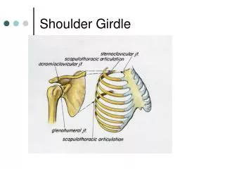
Shoulder Girdle Articulations • Scapulohumeral • Ball and Socket • Acromioclavicular • Gliding • Sternoclavicular • Double Gliding
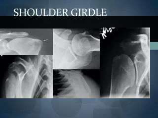
AP ProjectionShoulder (Anatomic Position, External Rotation)
AP ProjectionShoulder (Anatomic Position, External Rotation) • Greater tubercle and Humeral head in profile • Supraspinatus tendon insertion visualized
AP ProjectionShoulder (Neutral Rotation, palm against hip) • Greater Tubercle partially superimposing the humeral head • Posterior part of supraspinatus insertion demonstrated • Profiles calcific deposits not otherwise visualized
AP ProjectionShoulder (Internal Rotation, posterior hand against hip)
AP ProjectionShoulder (Internal Rotation, posterior hand against hip) • Lesser Tubercle in profile • Proximal humerus in true lateral position • Insertion site of subscapular tendon demonstrated
Transthoracic Lateral ProjectionShoulder (Lawrence Method) • What do you do if the patient cannot sufficiently elevate the unaffected shoulder?
Inferosuperior Axial ProjectionLawrence Method Degree of angulation of CR depends on abduction of arm
Inferosuperior Axial ProjectionLawrence Method Lesser Tubercle Humerus Coracoid Process Acromioclavicular Joint • Lesser Tubercle in profile • Coroacoid Process pointing anteriorly Acromion Scapulohumeral Joint
Superoinferior Axial ProjectionAlternative to Supine Lawrence Method • Place the patient in a chair at the end of the exam table and have them extend the shoulder over the table. • Shoulder should be over midpoint of IR • Tilt head away from IR • Humeral epicondyles should be vertical CR 5-15 degrees toward elbow
AP Axial ProjectionTrauma Shoulder • Demonstrates relationship of humeral head to the glenoid cavity • Useful in diagnosing posterior dislocation CR 35 degrees
Scapular YPA Oblique Projection • The position of the arm is unimportant because it does not change the relationship of the humeral head to the glenoid cavity
Scapular Y • Useful in demonstrating dislocations • Anterior Subcoracoid dislocation • Head beneath the coracoid process • Posterior Subacromial dislocation • Head projected beneath acromion process
AP Oblique ProjectionGlenoid Cavity(Grashy Method) • RPO / LPO Position • 35-45 degrees toward affected side • Scapula parallel with the plane of the IR • CR 2 in. medial and 2 in. inferior to superolateral border of the shoulder Open Glenoid Cavity in Profile
Intertubercular GrooveTangential Projection CR: 10-15 degrees posterior Hand supinated Profiles the intertubercular groove free from superimposition of the surrounding shoulder structures.
Acromioclavicular ArticulationsAP Projection: Bilateral • SID: 72 inches • Upright Position • With and Without weights • Demonstrates dislocation, separation, and the function of joints
Acromioclavicular ArticulationsAP Projection: Bilateral • What pathology does this image demonstrate? • How do you know a patient is not rotated or favoring the injured side?
Acromioclavicular ArticulationsAlexander Method • AC Joint and Clavicle projected above the Acromion CR 15 degrees cephalic
ClaviclePA Projection • What would be the advantage of doing a PA Projection?
AP Axial ProjectionLordotic Position • Thinner patients require more angulation to project the clavicle off of the scapula and ribs. • Which position is easier for the patient?
AP Axial ProjectionLordotic Position • How do you treat a fractured clavicle?
Scapula Lateral Projection • Patient flexes elbow and places hand on posterior thorax • Delineates the acromion and coracoid process • Adjust body of scapula to be perpendicular to the IR
Scapula Lateral Projection • Arm brought across the chest grasping opposite shoulder • Position of the arm determines what portion of the scapula will be superimposed by the humerus
ScapulaLateral Projection • Extending the arm upward demonstrates the body of the scapula best.
Shoulder Arthrography • Examination of a joint after the injection of contrast material that outlines soft tissue and joint structures. • The most common purpose of shoulder arthrography is to rule out bursitis