Download
1 / 28
280 likes | 476 Views
Lecture 05 Intro to Myology. Types of Muscle Skeletal – Cardiac – Smooth – What characterizes each? Where is each found?. Muscles pull on skeletal parts to ___________ Muscles maintain _______________ Muscle _____________ body cavities

E N D
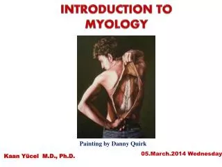
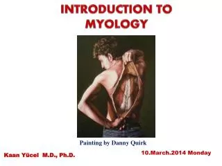
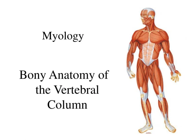
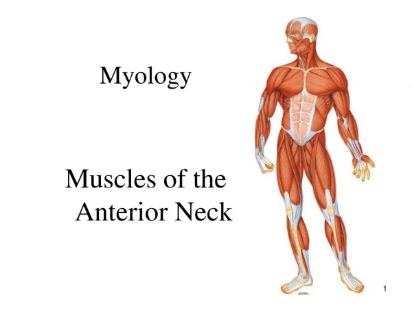
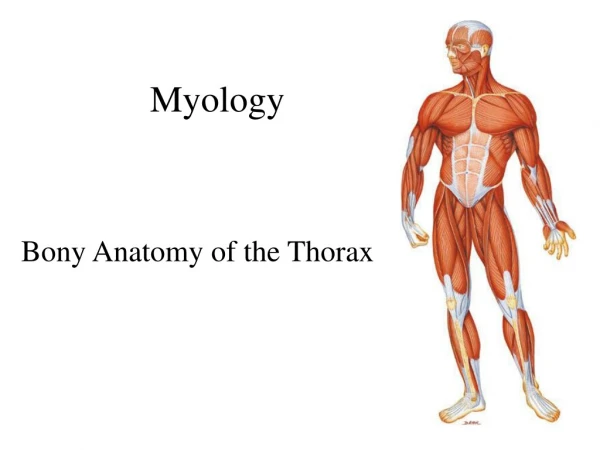
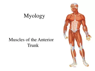
Lecture 05 Intro to Myology
Types of Muscle • Skeletal – • Cardiac – • Smooth – • What characterizes each? Where is each found?
Muscles pull on skeletal parts to ___________ Muscles maintain _______________ Muscle _____________ body cavities Muscles ___________ openings and thus _____________ entrance/exit of material from the body. Muscles produce body _________ and thus aid in maintenance of ____________.
Associations with Connective Tissue Coverings: • Superficial fascia • Deep fascia • Connective tissue associated with muscle- what do each of these layers cover? • epimysium • perimysium • endomysium • All contribute to formation of a tendon
Nerve and Blood Vessel Supply • Artery - Each muscle is supplied by an artery • Veins – 1-2 drain each muscle • Found in Epimyceum • Capillaries – ramify through endomyceum, 1 or 2 per muscle cell – wrap around muscle fiber
Skeletal Muscles are Controlled via Motor Neuron Motor unit = one somatic motor neuron & all the skeletal muscle cells (fibers) it stimulates One nerve cell supplies on average 150 muscle cells that all contract in unison. Total strength of a contraction # motor units activated Size of motor units
Tendon • Tie muscle to bone • Dense fibrous connective tissue • Little vascular supply • Slow to heal • May be enclosed within synovial sheath (wrist, ankles) • Aponeurosis – • Broad sheet of connective tissue • Epicranial aponeurosis
Connective Tissue Components aponeurosis tendon
Micro-Anatomy of Muscle Fibers: Sarco Muscle Sarcolemma-excitable membrane (T tubules) Sarcoplasm- glycogen, myoglobin, myofibrils Sarcoplasmic reticulum Stores calcium Triad- regulates calcium movement Sarcomere = contractile unit of myofibril
Myofibrils Specialized cytoskeletal elements contraction of muscle fiber 100’s – 1000’s / muscle fiber Organized into Sarcomeres ca. 10,000/myofibril Basic contractile unit Composed of: Contractile proteins Actin: thin filaments myosin: thick filaments Regulatory proteins Tropomyosin, troponin Structural proteins Titin, myomesin, dystrophin
Neural Control – via motor neuron Arrives at Neuromuscular Junction: motor end plate: sarcolemma at NMJ, location of neurotransmitter receptors Synaptic region – microscopic gap between neuron and muscle fiber Neurotransmitter (chemical) diffuses across gap to initiate contraction One NMJ/muscle fiber
Neuromuscular Junction (NMJ) or Synapse NMJ = myoneural junction end of axon nears the surface of a muscle fiber at its motor end plate region (remain separated by synaptic cleft or gap)
Structures of NMJ Region Synaptic end bulbs are swellings of axon terminals End bulbs contain synaptic vesicles filled with acetylcholine (ACh)= neurotransmitter
Muscle Contraction – Sliding Filament Theory Interaction between thick and thin filaments: Nerve impulse at neuromuscular junction causes release of Na ions following binding of acetylcholine on motor endplate Na ions cause release of Ca ions within muscle cells Ca ions make binding of thick and thin filaments possible ‘Heads’ on myosin (thick) filament bind at binding sites on Actin (thin) filament Binding causes myosin head to move, pulling actin filaments together
Muscle Cell Response THRESHOLD STIMULUS Muscle twitch (contraction) relaxation ALL OR NONE Response of a motor unit RECRUITMENT = strength of contraction from stimulation of additional fibers (activation of motor units) MUSCLE TONE = result of percentage of individual fibers contract and subsequently relax – thus lending support Muscle tone controlled by interaction of muscle spindle and muscle fibers in a motor unit TETANUS =smooth contraction from repeated stimulus - summation
Energy Required for Muscle Contraction ‘cocking of myosin head Pumping of Ca ions into sarcoplasmic reticulum Pumping of Na out of muscle fiber to neuromuscular cleft Energy as ATP Production of energy requires Oxygen Nutrient source (glucose from carbs)
Different Kinds of Muscle Fast fibers: (white fibers) High concentration of myofibrils Little reserve energy/replenishment capacity Rapid contraction Slow Fibers: (red fibers) Smaller diameter Replenish energy (ATP) as used Store oxygen in myoglobin within cell Dark color Intermediate Fibers – convert to fast fibers – intermediate properties
Variations in Skeletal Muscle Fibers Myoglobin, mitochondria and capillaries red muscle fibers more myoglobin, an oxygen-storing reddish pigment more capillaries and mitochondria white muscle fibers less myoglobin and less capillaries give fibers their pale color Contraction and relaxation speeds vary how fast myosin ATPase hydrolyzes ATP Resistance to fatigue different metabolic reactions used to generate ATP
Classification of Muscle Fibers Slow oxidative (slow-twitch) red in color (lots of mitochondria, myoglobin & blood vessels) prolonged, sustained contractions for maintaining posture Fast oxidative-glycolytic (fast-twitch A) red in color (lots of mitochondria, myoglobin & blood vessels) split ATP at very fast rate; used for walking and sprinting Fast glycolytic (fast-twitch B) white in color (few mitochondria & BV, low myoglobin) anaerobic movements for short duration; used for weight-lifting
Fiber Types within a Whole Muscle Most muscles contain a mixture of all three fiber types Proportions vary with the usual action of the muscle neck, back and leg muscles have a higher proportion of postural, slow oxidative fibers shoulder and arm muscles have a higher proportion of fast glycolytic fibers All fibers of any one motor unit are same. Different fibers are recruited as needed.