Download
1 / 55
550 likes | 691 Views
Nicholas J. Volpe, MD Tarry Professor and Chairman Department of Ophthalmology Feinberg School of Medicine Northwestern University. The Aging Eye : How to Keep Your Sight For Life. Strategies to Preserve Your Vision.

E N D
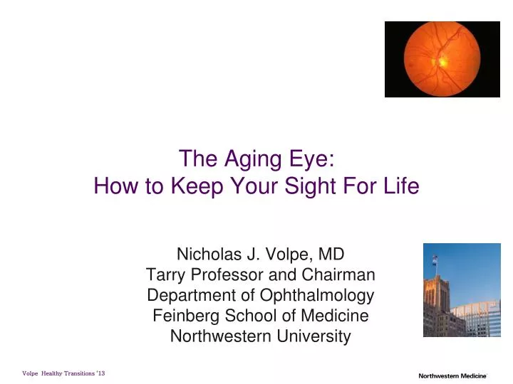
Nicholas J. Volpe, MD Tarry Professor and Chairman Department of Ophthalmology Feinberg School of Medicine Northwestern University The Aging Eye: How to Keep Your Sight For Life
Strategies to Preserve Your Vision • Prevention is our most potent tool in the quest to reduce disease (and healthcare costs) • Choose your parents well and stop aging!!! OR • Don’t Smoke • Wear Glasses that are UV protective • Safety glasses for high risk activities • Pay Attention to Nutrition and Vitamins • Don’t Ignore Symptoms • Get Regular Eye Examinations
Major Causes of Chronic Visual LossPreventable and Treatable Cataracts Glaucoma Macular degeneration Diabetic retinopathy Other Issues Dry eye Presbyopia near vision blurring
Protective Eye Ware • Avoid fireworks! • Always if you have poor vision in one eye • High risk activities • Racquet sports • Sawing • Drilling • Sawing • Working overhead • Any high speed tool • firearms
Stop Smoking • Clearly a risk factor for cataracts • 3X the risk • Clearly a risk factor for macular degeneration and its response to treatment
Nutrition • Healthy tear film • Macular degeneration • Fruits and Green Leafy Vegetables • Carotenoid pigments (lutein) accumulate in macula and prevent light damage • Omega fatty acids • Lutein and Zeaxanthin • Studied in AREDS 2 • Vitamins A,C, E
Regular Check Ups • Many diseases can be detected • Every 2-3 years from age 40-65 • Every 1-2 years after age 65 • More frequently with diabetes or family history of glaucoma or macula degeneration • Young adults, in the absence of symptoms, do not require routine examinations
Cataracts Expected if ≥ 60 years old 50% - 65 - 75 years old 70% > 75 years old Most common cause of decreased vision Symptoms Loss of acuity Difficulty with colors Glare at night Trouble reading small print Age Steroids (PSC) Trauma Inflammation Diabetes Other drugs
Subcapsular cataract Anterior Posterior
Nuclear cataract Progression • Increasing nuclear opacification • Exaggeration of normal nuclear • ageing change • Causes increasing myopia • Initially yellow then brown
Classification according to maturity Immature Mature Hypermature Morgagnian
Drugs Systemic or topical steroids Chlorpromazine - initially posterior subcapsular - central, anterior capsular granules Other drugs • Long-acting miotics • Amiodarone • Busulphan
Cataract Surgery Outpatient Very successful > 95% Almost all with intraocular lenses Most common surgical procedure in U.S. >1.4 million/year Most successful surgical intervention Complications uncommon sight threatening IOL technology continues to evolve for astigmatic correction and presbyopia Newest modality is femtosecondlaser
Cataract Prevention • Smoking cessation • Reduces Vitamin C in the eye • Vitamin C levels are high in the eye and this helps remove prooxidants • Fruits and vegetables • 5 fold decrease at 3-4 servings per day • Regular alcohol consumption increases risk of cataract • Steroids and inflammatory conditions are risks for cataracts • Obesity and radiation
Ultraviolet Light • Cataracts and Macular Degeneration • Cataracts much more prevalent in equatorial climate • AMD more common in light eyes • Same rules as sun tan lotions • if you might tan or burn you should be wearing sunglasses • 10-30% transmission of light • Wide brimmed hat • Also water, sand and snow • Polarized not necessary but will cut glare • Don’t assume expensive is UVA and B protective • Test lens quality and fit to ensure successful use
Age- Related Macular Degeneration Age-related macular degeneration (AMD) is the most common cause of severe, irreversible vision loss in older Americans and Europeans. (AMD Alliance International 2008; Ferris et al. 1984; National Society to Prevent Blindness 1980). Worldwide, AMD disease affects 25-30 million people. Etiology is complex and poorly understood Free-radical mediated damage to the photoreceptors and the RPE may disrupt the transport of metabolites from photoreceptors to choroidal capilaries Angiogenesis is a feature of neovascular AMD AMD may be associated with a systemic vascular disorder Genetic and environmental factors Variation in the complement factor H gene
Free radicals and antioxidants in CNV light free radical production damage blocked by antioxidants photoreceptors and RPE damage
AMD Risk Factors Gender ♀ > ♂ Race/Ethnicity Smoking Family History Atherosclerosis Hypertension Symptoms early = None, mild distortion late = acute loss of vision
Atrophic AMD Progression Initially drusen and non-specific RPE changes Late RPE (geographic) atrophy
Atrophic AMD Fluorescein angiogram Management Hyperfluorescence from RPE window defect Low-vision aids if appropriate
Pathophysiology: Penetration of Bruch’s Membrane schematic fundus photograph New blood vessels penetrate Bruch’s membrane
Choroidal Neovascularization (CNV) • Less common than atrophic AMD but more serious • Metamorphopsia is initial symptom • Many lesions are not visible clinically Suspicious clinical signs Subretinal blood or lipid Gray-yellow subretinal lesion with fluid
Current Status of Therapiesfor CNV Antiangiogenic therapy Lucentis, Avastin, Macugen CATT trial (Avastin vs Lucentis) Photodynamic therapy with verteporfin Steroids Thermal Laser
Treatment for Dry AMD -Age-related Eye Disease Study (AREDS) –role of antioxidants vitamin E, 400 IU vitamin C, 500 mg beta carotene, 15 mg (approximately 25,000 IU Vitamin A) zinc 80 mg as zinc oxide copper, 2 mg, as cupric oxide Copper should be taken with zinc, because high-dose zinc is associated with copper deficiency.
Established Age Related Macular Degeneration • Use Amsler Grid to monitor central vision • AREDS-OccuvitePreservision • B carotene vs. Lutein and Zeaxanthin (AREDS 2) • Vitamin C • Vitamin E • Zinc Oxide (?necessary and ? Stomach upset) • Copper • NB: No beta carotene for smokers and others at risk for lung cancer • Others??? Lutein Eyes, PhoVision, Perspective, Ocu-force
AREDS Results Recommendations Evaluation: Persons over 55 years old receive a dilated eye exam to assess risk of advanced AMD. Contraindications to Treatment: Smokers and ex-smokers should not use beta carotene, because previous studies have suggested an association with lung cancer and beta carotene in smokers. There were no benefits from treatment shown in the AREDS for patients with no AMD (Category 1) and early AMD (Category 2).
AREDS 2 • Adding omega 3’s did not help • Taking away B Carotene did not hurt and lutein and zexanthine may have been a bit more protective • Reducing zinc dose did not hurt and less side effects • No prevention of cataracts
Diabetic Retinopathy • most common cause of • new blindness among adults 20-64 yo • Blindness in working adults • affects over 5.3 million Americans age >18 (2.5% of this population) • Prevention- worse in HTN, obesity, renal failure, hyperlipidema, smoking, anemia, pregnancy and POOR glycemic control
Clinical Findings in NPDR Microaneurysms Earliest clinical sign of diabetic retinopathy Appear as small red dots in the superficial retinal layers Rupture produces blot/flame hemorrhages
Macular Edema (CSME) Leading cause of visual impairment in patients with diabetes
Macular Edema Treatments ETDRS focal laser surgery for CSME reduces the incidence of moderate visual loss (doubling of visual angle or roughly a 2-line visual loss) from 30% to 15% over a 3-year period Steroids -peri-ocular -intraocular Anti-VEGF agents
Ischemic diabetic maculopathy • Macula appears relatively normal • Capillary non-perfusion on FA • Poor visual acuity • Treatment not appropriate
PDR Proliferation of new blood vessels due to ischemia NVD Disc NVE Elsewhere NVI Iris NVA Angle
PDR - cont. Treatment Options Pan-retinal photocoagulation Peripheral Retinal Cryotherapy Vitrectomy Anti-VEGF
Retinopathy Screening Type 1 diabetes - screen within 3-5 years of diagnosis after age 101 Type 2 diabetes - screen at time of diagnosis1 Pregnancy - women with preexisting diabetes should be screened prior to conception and during first trimester1 Follow-up depends on severity of disease
Diabetic Eye Care • Like glaucoma, you will NOT HAVE SYMPTOMS UNTIL IT IS TOO LATE! • 95-100% treatable with early detection • Regular eye exams at 6 or 12 month interval depending on what MD sees • Bleeding, swelling and growth of blood vessels • Diabetes control (Hemoglobin A1c) is the most important way to reduce your risk • High blood pressure is a risk • Diet and exercise
Glaucoma Optic nerve 1.2 million nerve fibers Ganglion cells in retina exit to brain as optic nerve
Definition of Glaucoma • A group of optic neuropathies in which retinal ganglion cells die by apoptosis with resultant optic disc cupping and characteristic visual field deficits • Optic neuropathy • Retinal ganglion cell apoptosis • Optic disc cupping or excavation • Loss of visual function -IOP is too high for the nerve??? • Most common cause blindness: • African-Americans COMPLETE/TOTAL BLINDNESS
Glaucoma Loss of visual field Site of visual field loss corresponds to area of damage on optic disc, e.g., “cupping”
Classification of the Glaucomas • RISK FACTORS • IOP 21 mm Hg • Family history • Risk is increased by x2 if parent has POAG • Risk is increased x4 if sibling has POAG • African American • C:D ratio > 0.5 • Asymmetric cupping • Myopia • Diabetes • Age > 75 years • Open-angle glaucomas (90%) • Angle-closure glaucomas (10%) • Primary glaucomas • Secondary glaucomas
Angle Closure Glaucoma Acute pain, redness, tearing Associated with dilation of pupil Natural (e.g., movie theater) Pharmacologic Nausea & vomiting often in ER with “acute abdomen” Risk factor of narrow angle can be detected on screening exam (esp hyperope) and prophylactic iridotomy is preventative of attack in 100%
Acute angle-closure glaucoma Signs • Ciliary injection • Complete angle closure • Severe corneal edema • Shallow anterior • chamber • Dilated, unreactive, • vertically oval pupil Medical rx to lower IOP, followed by laser (Yag) iridotomy
Primary Open-Angle Glaucoma • The most prevalent type of glaucoma in the United States • Elevated intraocular pressure is not part of the diagnostic criteria • 25% of patients with primary open-angle glaucoma in the US have normal intraocular pressure • Asymptomatic • Some loss of visual field • Most common type • Familial, bilateral • “Sneak thief of sight”
Primary Open-Angle Glaucoma • Evidence that IOP reduction is beneficial • Collaborative Normal-Tension Glaucoma Study (CNTGS) • Advanced Glaucoma Intervention Study (AGIS) • Early Manifest Glaucoma Study (EMGT) • 25% IOP reduction RoP 62% to 45% at a median of 6 years. • Ocular Hypertension Treatment Study (OHTS)
Treatment for POAG • Lower the IOP • Medical therapy • Prostaglandin , B-blockers,Sypathomimetics, Carbonic-anyhrase inhibitors • Laser surgery (ALT, SLT) • Incisional surgery (Trab, shunt)