Download
1 / 60
730 likes | 1.49k Views
Liver regeneration- principles, measurement and status. Perceptor Prof. S K Acharya. Introduction. Liver possesses an ability to regenerate following partial resection until it attains its original size

E N D
Liver regeneration- principles, measurement and status Perceptor Prof. S K Acharya
Introduction • Liver possesses an ability to regenerate following partial resection until it attains its original size • Clinically important- this can be an alternative to liver transplantation
Introduction • Regeneration means the reconstitution of a structure that has been excised. Eg. complete re-growth of the limb of a newt or tail of lizard • In c/o liver it is actually compensatory hyperplasia rather than true regeneration • Total liver mass rather than the lobulated anatomic configuration is restored
Introduction • In normal liver fewer than 1 in 10,000 hepatocytes undergo mitosis at any given time, they remain in Go • Normal hepatocytes respond poorly to mitogens like TGF, EGF & HGF • Liver possesses a unique capacity to replace tissue mass after injury or loss of liver mass • Regenerative capacity is insufficient - CVH or alcoholism- cirrhosis
Experimental models • Model to study liver regeneration is removal of 2/3 of the liver of rats/mice a technique known as 2/3 PHx • Liver cells proliferate - original liver mass is restored within 5-7 days • No massive inflammation, fibrosis or scarring Bucher NL. Regeneration of Mammalian Liver.Int. Rev. Cytol.1963;15: 245–300
Rodent model • Hepatocytes are the first to enter the cell cycle • Undergo 2 rounds of cell division within 2–3 days • Proliferation of hepatic stellate cells, Kupffer cells and biliary epithelial cells • Angiogenesis (endothelial cells) occur to re-establish the liver vasculature Bucher NL. Regeneration of Mammalian Liver.Int. Rev. Cytol.1963;15: 245–300
Rodent model • The advent of gene knock out mice - various specific molecules and dissection of pathways • No single GM mouse model demonstrates 100% mortality and a complete blockage of both DNA replication and cell proliferation • No single gene can be considered “essential” for liver regeneration
Check points in cell division Hepatocytes in the normal liver are quiescent (Go phase) and respond minimally to in vivo mitogens such as TGF EGF and HGF These mitogens can induce replication following infusion of TNF-a
Timing of Regeneration • In rat, DNA synthesis peaks at 24 hours after PH, when approximately 35% of hepatocytes are in cell cycle • Observed- circadian clock controls the G2/M transition • G2-M transition always occur at the same time of day Matsuo T, Yamaguchi S, Mitsui S, Emi A, Shimoda F, Okamura H. Control mechanism of the circadian clock for timing of cell division in vivo. Science 2003;302:255-25
Timing of Regeneration • Mammals- Circadian timing system controlled by pacemaker in the suprachiasmatic nucleus of the brain • Synchronizes subsidiary circadian oscillators in peripheral cell types including hepatocytes Matsuo T. Control mechanism of the circadian clock for timing of cell division in vivo. Science 2003;302:255-25
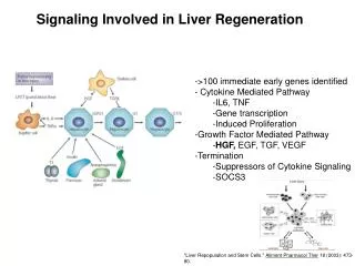
Immediate Early Genes • Activated almost immediately (within minutes) after partial hepatectomy in mice • 70 immediate early genes and more • Proto-oncogenes c-fos, c-jun, c-myc, and c-ets • Transcription factors- NFκB , STAT3, IGF binding protein-1 etc
NFΚB AND IMMEDIATE EARLY GENES • Quiescent liver NFκB in the cytosol bound to IkB • Binding of TNF-a to TNF-R1- IkBkinase- Phosphrln of IκB • Free NFkBTranslocates to nucleus • Transcription of IL-6 • Activation of STAT3
Delayed Early Genes • Transcribed after the immediate early gene response • Expression occurs during later phases • Depends on protein synthesis; Tr Factors • HRS/SRp40 (splicing factor and modulator of alternative splicing of RNA transcripts) and the anti apoptotic gene bcl-x • Pro-apoptotic genes BAK, BAD, BAX - down-regulated
Cyclins and CDKs • Family of proteins that control the progression of cells through the cell cycle by activating Cdk • CDk- ser/threokinases inactive • Cyclin attachment activation • All CDKs perform phosphorylation of target proteins
Cyclins and CDKs • G1 phase; CDKs catalyze the phosphorylation of the pRb and cause its dissociation from the E2F family of proteins • This eliminates the repression of gene expression by pRB • Cyclin D1 also may sequester the cell cycle inhibitor p27with advancement in cell cycle
Integration of Cytokines and Growth Factors in Liver Regeneration • Priming • 0-5 hrs • Reversible phase in liver regeneration, G0 - G1 phase • Cytokines, eg. TNF-α and IL-6 • NFκB and STAT3 are activated • Sensitize hepatocytes to growth factors
Integration of Cytokines and Growth Factors in Liver Regeneration • Progression • Requires HGF (mesenchymal cells) and TGF-α (hepatocyte) & cyclins • During the progression phase, the cells move past the restriction point in G1 to S and beyond • GH, PTH, T3 are permissive for liver regeneration • Insulin and norepinephrine are adjuvant factors • HGF and c-met • Sources of HGF in the liver are Kupffer cells and HSC • HGF binding to of c-met activates tyrosine kinase and thus initiates a signal transduction pathway
Summary; the sequence of signals that leads to liver regeneration
Mitogenic Signals Associated With Initiation of Liver Regeneration • Quiescent hepatocytes express a variety of GF receptors; PDGF, VEGF, FGF receptors • Only mitogens for hepatocytes in media are HGF and ligands of the EGFR ; EGF, TGFα, AR, HBEGF, etc • Direct mitogens- induce a strong mitogenic response in hepatocytes • HGF, EGF, and TGFα also induce hepatocyte proliferation when injected into mice and rats
Mechanisms of regeneration in ALF • STAT3 translocates to the nucleus with induction of transcription of target genes • Go- G1; driven by HGF and EGF receptor ligand family • Serum levels of HGF have been found to be markedly elevated in the serum of adults and children in ALF Changes in serum levels of hepatocyte growth factor in patients undergoing adult-to-adult living-donor liver transplantation. Transplantation 2003;76:1769–1770
Oval cells • HPCs- generated from the biliary compartment in response to hepatic injury • Canals of herring, or in periductular situation • Capable of generating cholangiocytes or hepatocytes • Growth factors that stimulate oval cell proliferation are similar to those that stimulate hepatocyte replication
Oval cells • Murine liver +ve for • Immature markers - α-fetoprotein • Mature hepatic markers (e.g., albumin) • Biliary markers (e.g., cytokeratin-19) • c-kit, CD34, Ov6, CK7, cgnn A + • Oval cells and hepatocytes require signaling through TNF-R1; but not simultaneously • TGF beta - oval cell replication is less affected
Oval cells • HPCs proliferate in the portal zone • “Streaming liver hypothesis” – migrate toward the central vein in the liver lobules as daughter hepatocytes • Proven with Mt DNA mutation tracking - in the human liver as well as in regenerative nodules in cirrhosis
Bone Marrow Stem Cells inLiver Disease • Stem cells - totipotent, pluripotent, multipotent, or unipotent • 2 endogenous populations of stem cells in liver- Hepatocytes and hepatic oval cells • Hepatocytes ; able to self-renew limitlessly often play the principal role in liver regeneration
Hematopoetic stem cells • Liver - location of hematopoiesis in fetus • ?Hematopoetic stem cells remain behind to form HSC • HSCs can share cell surface markers associated with hematopoietic stem cells such as CD34, Thy-1 and c-kit
Can Hematopoietic Cells Generate Hepatocytes? • Data from Murine Models • BM-HSC have been demonstrated to repopulate liver and give rise to functional hepatocytes • BM HSC derived hepatocytes- arise from cell fusion of donor HSC and recipient hepatocytes
Can Hematopoietic Cells Generate Hepatocytes? • Allogeneic bone marrow cells transplanted into lethally irradiated mice- generate hepatocyte-like cells in the liver at very low frequency (1/ 10,000)
Clinical Data from Sex-Mismatched BM Transplants • Microchimerism; Presence of cells that originate from another individual and are genetically distinct from the cells of the host individual • Frequency of BMHSC derived hepatocytes varied between <1% and 8% in sex mismatched individuals • Higher frequencies in pediatric liver allografts • Microsatellite analyses- higher percentage of chimerism in comparison to Y chromosome evaluation by FISH
Liver regeneration in acute severe liverimpairment: a clinicopathologicalcorrelation study • N = Liver Bx samples of 74 patients with ALF/SALF • Etiology; acute HAV + drug toxicity (n 3), acute HBV (n 10), drug induced (n 21) and cryptogenic (n 40) • Mean age: 43+17 years • M/F: 28/ 46 • IHC for CK7/CK19 – HPCs activation/differentiation • Ki 67/P21 - proliferative activity/proliferation arrest • H & E - hepatocyte loss AezamKatoonizadeh. Liver international 2006
Liver regeneration in acute severe liver impairment • Oval cell activation requires 50% loss of hepatocytes, a/w significant decrease in the proliferative activity of remaining hepatocytes • Associated with poor prognosis AezamKatoonizadeh. Liver international 2006
Mesenchymal Stem Cells as a Source forLiver Regeneration • Multipotentstromal cells that can differentiate into osteoblasts, chondrocytes , adipocytes • In vitro; Differentiate into hepatocyte like cells and hepatic epithelial cells • Umbilical cord blood, wharton’sgelly, adipose tissue • Hypoimmunogenic and create immuno suppressive microenvironment • 1 gram of fat > no of stem cells 1 gram of marrow
Mesenchymal Stem Cells as a Source forLiver Regeneration • In vitro functional assays- hepatocyte characteristics • albumin production • glycogen storage • urea secretion • uptake of LDL • Studies in humans hindered by • Safety concerns • Lack of molecular data • Immunological mismatch Gut,vol.58,no.4
Hepatocyte transplantation • Involves the transfer of normal hepatocytes into diseased liver • Isolated hepatocytes injected into splenic artery or directly into the portal vein
Hepatocyte transplantation • Host liver architecture remains intact following the integration of hepatocytes in liver cords • Transplantation is metabolically less stressful than whole liver transplants • Consequences of graft loss are much less severe • Does not interfere with subsequent liver transplantation or gene therapy
Murine studies in Hepatocyte transplantation • Murine model- u TPA over producing cells • 10 4 liver cells • replace more than 80% of the liver • donor hepatocytes capable of at least 12 rounds of cell division Replacement of diseased mouse liver by hepatic cell transplantation. Science.263:1149–1152
Murine studies in Hepatocyte transplantation • Intraperitoneal injections of hepatocytes into rats with fulminant hepatic failure induced by D galactosamine • improved survival • donor liver cells did not repopulate the liver Cellular transplantation in the treatment of experimental hepatic failure. Science. 1980. 210:901–903
Methods for Assessing Liver Regeneration • Need to accurately document the extent of hepatic regenerative activity • Patients with liver disease • Partial hepatic resections • Receiving interventions designed to stimulate hepatic regeneration
Methods for Assessing Liver Regeneration • Ideally • Non invasive, in-vivo • Measuring only specific phase of cell cycle • Sensitive and Specific
Methods for Assessing Liver Regeneration • Tissue determination • Liver weight • CT/MRI- estimated weight (in grams) equals 0.81 × hepatic volume in ml +372
Mitosis measurement • Incubating hepatocytes with potassium chloride and alcohol • Centrifugation • McNeill's stain • No of mitotic figures per 1000 cells per hpf • No of mitotic figures available for counting is relatively limited
DNA synthesis • Thymidinelabelling index • TK rate limiting enzyme for DNA synthesis • Phosphorylatesthymidine ; subsequent incorporation into the DNA • Liver biopsy tissues or isolated hepatocytes are incubated with radiolabelledthymidine for 1 h prior to measuring radioactivity • Standardized and reproducible marker of hepatic regenerative activity