Download
1 / 23
230 likes | 364 Views
Explore the vital role of blood as a fluid connective tissue carrying nutrients and waste products. Learn about the components, microscopic features, and functions of red blood cells, leukocytes, and platelets in light and electron microscopy.

E N D
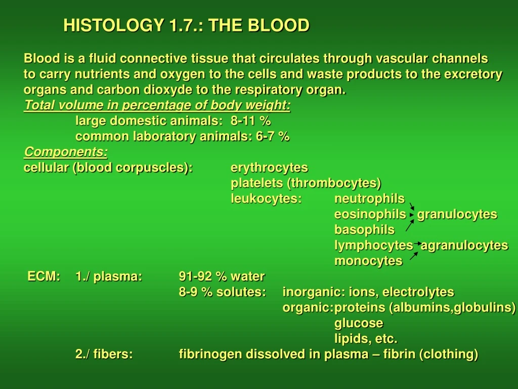
HISTOLOGY 1.7.: THE BLOOD Blood is a fluid connective tissue that circulates through vascular channels to carry nutrients and oxygen to the cells and waste products to the excretory organs and carbon dioxyde to the respiratory organ. Total volume in percentage of body weight: large domestic animals: 8-11 % common laboratory animals: 6-7 % Components: cellular (blood corpuscles): erythrocytes platelets (thrombocytes) leukocytes: neutrophils eosinophils granulocytes basophils lymphocytes agranulocytes monocytes ECM: 1./ plasma: 91-92 % water 8-9 % solutes: inorganic: ions, electrolytes organic: proteins (albumins,globulins) glucose lipids, etc. 2./ fibers: fibrinogen dissolved in plasma – fibrin (clothing)
Microscopic preparation of blood for light microscopic studies An evaluation is often needed to assess general health, or to diagnose haematologic and some other diseases. Blood test is used for this purpose. For light microscopic observation one drop of blood is enough spread onto the surface a slide. The blood film is air-dried, fixed in methanol and stained with Giemsa
Blood corpuscles • Erythrocytes (red blood cells) • Non-nucleated biconcave discs: dog, cow, sheep. • Shallow concavity: horse, cat • Flat shape: pig, goat • Size and number also varies among species SEM image of blood LM image of a blood smear
1.RED BLOOD CELLS (ERYTHROCYTES) Size: 7 mm the largest (in dog) 4.1 mm the smallest (in goat) Number in 1 mm3 of blood: in the range of N x 106 (depending on size) Origin: red bone marrow Life span: some months only (120 days) Destruction: in the spleen Poikilocytosis: variation in shape (goat, deer) Rouleaux formation: adherence to each other, forming long chains Howell-Jolly bodies: small, round, pyknotic DNA fragments (remnants of the nucleus) Reticulocyte: immature red blood cell with residual ribosomes
Rouleaux formation Howell-Jolly bodies (arrows) Poikilocytosis Reticulocyte (arrow)
Fine structure of erythrocytes: Mature erythrocyte lacks nucleus and cell organelles. It is a membrane-bound container of hemoglobin (33-36 %) some enzymes and electrolytes in water. Biconcave shape is maintained by spectrin at the internal surface of the membrane TEM Main function of the erythrocytes: to carry oxygen to the cells and to carry carbon dioxyde from the cells to the respiratory organ
Molecular structure of hemoglobin Heme-part
2. Leukocytes (white blood cells) (5000-9000/mm3): 2.1. Polymorphonuclear granulocytes: 2.1.1. Neutrophils (55-60 %) 2.1.2. Eosinophils (1-3 %) 2.1.3. Basophils (0-0.7 %) 2.2. Mononuclear agranulocytes 2.2.1. Lymphocytes (25-33%) 2.2.2. Monocytes (3-7 %) 2.1.1. 2.2.1.
2.1.1. Neutrophils: The most segmented nuclei (4-5 lobes) Size: 10-12 mm Amount: 3000-6000/ml Moderately stained pale granules Function: phagocytosis (microphages) Origin: red bone marrow Life span: some hours (7-8)
2.1.1. Neutrophils: • Ultrastructure: • Two types of granules: • Specific: lysosyme, • lactoferritin, alkaline • phosphatase • Azurophilic: lysosomal • enzymes (acidic phos- • phatase, peroxidase, • etc.) • Neutrophils are able to • migrate through capillary walls (see the image) to the site of bacterial • infection. • There they phagocyte the bacteria. • Pus: phagocyted bacteria and hundreds of thousands dead neutrophils. Endothelial cell
2.1.2. Eosinophils: Bilobed nuclei (spectacle form in human) Bright red eosinophilic granules (species-specific) Size: 10-12 mm Amount: 300/ml Origin: red bone marrow Life span: 8-12 days Function: Control of allergic reactions Moderate phagocytotic activity Defense against parasitic worms
2.1.2. Eosinophils: Fine structure: Specific granules are heterogeneous from species to species (various crystal structures inside). Content of specific granules: lysosomal hydrolases, major basophilic protein MBP, eosinophilic cationic protein ECP, eosinophil-derived neurotoxin, antihistamin, slow reacting substance SRS) TEM
2.1.3. Basophils: U- or kidney-shaped nucleus Large purple granules Size: 10 mm Amount: very rare Origin: red bone marrow Function: They share common features with the CT mast cells. Their granules contain histamine (allergic reactions, anaphylaxic shock), heparin (anticoagulant). Human
2.2.1. Lymphocytes Large nucleus, thin cytoplasmic rim Size: 7-12 mm (large-medium-small) Origin: red bone marrow No stained granules in the cytoplasm (except small azurophilic ones) Types: B- and T-lymphocytes (morphologically not distinguishable) null-cells (somewhat smaller size) Function: defence - immunity
2.2.2. Monocytes: Large kidney-shaped eccentric nucleus No granules in the cytoplasm except azurophilic small granules Size: up to 17 mm Origin: red bone marrow Function: 1-2 days circulation in the blood, then enter the CT and trasform into macrophages (see there) TEM
3. Platelets (thrombocytes): Minute colorless anucleate corpuscles Thin biconvex discs Size: 2-3 mm Number: 150.000-300.000/ml Origin: megakaryocytes Function: blood clothing TEM TEM LM
Blood clothing Thromboplastin Prothrombin Thrombin Fibrinogen Fibrin Ca ions are also needed for the process
The avian blood: Erythrocytes are nucleated ovoid cells Thrombocytes are nucleated ovoid cells Granulocytes are called heterophils (arrows) Please note: all the submammalian vertebrate animals (fish, amphibians, reptiles) have similar ovoid nucleated red blood cells