Chapter 4: Cellular Form and Function
Chapter 4: Cellular Form and Function. Development of the cell theory: Hooke in 1663, observed cork (plant): named the cell Schwann in 1800’s states: all animals are made of cells Pasteur’s work with bacteria ~ 1860 disproved idea of spontaneous generation (living things from nonliving)

Chapter 4: Cellular Form and Function
E N D
Presentation Transcript
Chapter 4: Cellular Form and Function Development of the cell theory: • Hooke in 1663, observed cork (plant): named the cell • Schwann in 1800’s states: all animals are made of cells • Pasteur’s work with bacteria ~ 1860 disproved idea of spontaneous generation (living things from nonliving) • Modern cell theory emerged by 1900
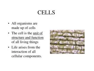
Modern Cell Theory • All organisms composed of cells and cell products. • A cell is the simplest structural and functional unit of life. There are no smaller subdivisions of a cell or organism that, in themselves, are alive. • An organism’s structure and all of its functions are ultimately due to the activities of its cells. • Cells come only from preexisting cells, not from nonliving matter. All life, therefore, traces its ancestry to the same original cells. • Because of this common ancestry, the cells of all species have many fundamental similarities in their chemical composition and metabolic mechanisms.
Cell Shapes • thin, flat, angular contours • round to oval • irregular angular shapes, > 4 sides • disc shaped
Cell Shapes 2 • squarish • thick middle, tapered ends • taller than wide • long, slender Stellate • nerve cells have extensions, look starlike
Epithelial Cell Surfaces • Epithelial cells line organ surfaces • Basal surface • cell rests on this lower surface • Lateral surface • the sides of the cell • Apical surface • exposed upper surface
Cell Size • Human cell size • most range from 10 - 15 µm • egg cells (very large)100 µm diameter, visible to naked eye • nerve cell over 1 meter, muscle cell up to 30 cm, (too slender to be seen) • Limitations on cell size • as cell enlarges, volume increases faster than surface area so the need for increased nutrients and waste removal exceeds ability of membrane surface to exchange
Evolving Perspective on Cells • Early study with light microscope revealed • surface membrane, nucleus and cytoplasm • Electron microscopes have much higher resolution and revealed much greater details, such as the cell ultrastructure of the cytoplasm • fibers, passageways and compartments, and organelles surrounded by cytosol (a clear gelatinous component also called intracellular fluid)
Plasma Membrane • Defines cell boundaries • Controls interactions with other cells • Controls passage of materials in and out of cell • Appears as pair of dark parallel lines around cell (viewed with the electron microscope) • intracellular face - side faces cytoplasm • extracellular face - side faces outwards • Structure described by fluid-mosaic theory • arrangement of mobile globular proteins embedded in an oily film of phospholipids
Membrane Lipids • Lipids constitute • 90 to 99% of the plasma membrane • Glycolipids • 5% of the lipids, found only on extracellular face, contribute to glycocalyx
Membrane Lipids 2 • Cholesterol • 20% of the lipids, affects membrane fluidity (low conc.. more rigid, high conc.. more fluid) • Phospholipid bilayer • 75% of the lipids, with hydrophilic heads (phosphate) on each side and hydrophobic tails in the center • motion of these molecules creates membrane fluidity, an important quality that allows self repair
Membrane Proteins • Proteins constitute • only 1 to 10% of the plasma membrane, but they are larger and account for half its weight • Integral (transmembrane) proteins • pass through membrane, have hydrophobic regions embedded in phospholipid bilayer and hydrophilic regions extending into intra- and extracellular fluids • most are glycoproteins, conjugated with oligosaccharides on the extracellular side of membrane
Membrane Proteins 2 • Integral proteins (cont.) • may cross the plasma membrane once or multiple times • Peripheral proteins • adhere to intracellular surface of membrane • anchors integral proteins to cytoskeleton
Membrane Protein Functions • Receptors • Second messenger systems • Enzymes • Channel proteins • Carriers and pumps • Motor molecules • Cell-identity markers • Cell-adhesion molecules
Protein Functions - Receptors • Cells communicate with chemical signals that cannot enter target cells • Receptors bind these messengers (hormones, neurotransmitters) • Each receptor is usually specific for one messenger
Second Messenger System • A messenger (epinephrine) binds to a receptor 1 • Receptor releases a G protein 2 • G protein binds to an enzyme, adenylate cyclase, which converts ATP to cAMP, the 2nd messenger 3 • cAMP activates a kinase 4 • Kinases add Pi, activates or inactivates other enzymes
Enzymes in Plasma Membrane • Break down chemical messengers to stop their effects • Final stages of starch and protein digestion in small intestine • Involved in producing second messengers (cAMP)
Protein Functions - Channel Proteins • Formed by integral proteins • Channels are constantly open, allow water and hydrophilic solutes in and out
Protein Functions - Channel Proteins 2 • Gates open to three type of stimulants • ligand-regulated gates: bind to chemical messenger • voltage-regulated gates: potential changes across plasma membrane • mechanically regulated gates:physical stress such as stretch and pressure • Gates control passage of electrolytes so are important in nerve signals and muscle contraction
Protein Functions - Motor Molecules • A filamentous protein that arises deep in the cytoplasm and pulls on membrane proteins causing movement: • within a cell (organelles) • of a cell (WBC’s) • shape of cell (cell division, phagocytosis)
Protein Functions - Carriers • Integral proteins that bind to solutes and transfer them across membrane • Carriers that consume ATP are called pumps
Protein Functions - Cell-identity Markers • Glycoproteins contribute to the glycocalyx, a surface coating that acts as a cell’s identity tag
Protein Functions - Cell-adhesion Molecules • Membrane proteins that adhere cells together and to extracellular material
Glycocalyx • Surface of animal cells • CHO moieties of membrane glycoproteins and glycolipids that retains a film of water • Functions • immune response to infection and cancer • basis of tissue transplant compatibility • cellular uptake of water, dissolved solutes • assists in cell adhesion, fertilization and embryonic development
Microvilli • Structure • extensions of plasma membrane (1-2m) that increase surface area for absorptive cells (by 15- 40x in intestine, kidney) • Brush border • on some cells, they are very dense and appear as a fringe on apical cell surface • Milking action • protein filaments (actin) attach from the tip of microvillus to its base, anchors to a protein mesh in the cytoplasm called the terminal web and can shorten pushing absorbed contents into cell
Cilia • Hairlike processes 7-10m long, 50-200 on cell surface move mucus, egg cells • Covered by saline layer created by chloride pumps • Cilia beat in waves, sequential power strokes followed by recovery strokes
Cilia 2 • Axoneme has a 9+2 structure of microtubules • 2 central microtubules stop at cell surface • 9 pairs of peripheral microtubules continue into cell as a basal body that acts as an anchor • dynein (motor protein) arms on one pair of peripheral microtubules crawls up adjacent pair bending cilia • Sensory cells • some cilia lose motility and are involved in vision, smell, hearing and balance
Flagella • Long whiplike structure that has an axoneme identical to that of a cilium • Only functional flagellum in humans is the tail of the sperm
Nucleus • Largest organelle • Nuclear envelope surrounds nucleus with two unit membranes • Contains DNA, the genetic program for a cell’s structure and function
Endoplasmic Reticulum • Rough ER • extensive sheets of parallel unit membranes with cisternae between them and covered with ribosomes, continuous with nuclear envelope • function in protein synthesis and production of cell membranes • Smooth ER • lack ribosomes, cisternae more tubular and branch more extensively, continuous with rough ER • function in lipid synthesis, detoxification, calcium storage
Ribosomes • Small dark granules of protein and RNA free in cytosol or on surface of rough ER • Interpret the genetic code and synthesize polypeptides
Golgi Complex • Synthesizes CHO’s, processes proteins from RER and packages them into golgi vesicles • Golgi vesicles • irregular sacs near golgi complex that bud off cisternae • some become lysosomes, some fuse with plasma membrane and some become secretory vesicles • Secretory vesicles • store a cell product for later release
Lysosomes • Package of enzymes in a single unit membrane, variable in shape • Functions • intracellular digestion - hydrolyze proteins, nucleic acids, complex carbohydrates, phospholipids and other substrates • autophagy - the digestion of worn out organelles and mitochondrion • autolysis - programmed cell death • glucose mobilization - lysosomes in liver cells break down glycogen
Peroxisomes • Appear similar to lysosomes, lighter in color • Abundant in liver and kidney • Function • neutralize free radicals • produce H2O2 in process of alcohol detoxification and killing bacteria • break down excess H2O2 with the enzyme catalase • break down fatty acids into acetyl groups
Mitochondrion • Double unit membrane • Inner membrane contains folds called cristae • ATP synthesized by enzymes on cristae from energy extracted from organic compounds • Space between cristae called the matrix • contains ribosomes and small, circular DNA (mitochondrial DNA) • Reproduce independently of cell and live for 10 days
Centrioles • Short cylindrical assembly of microtubules, arranged in nine groups of three microtubules each • Two centrioles, perpendicular to each other, lie near the nucleus in an area called the centrosome • these play a role in cell division • Other single centrioles migrate to plasma membrane forming basal bodies of cilia or flagella • two microtubules of each triplet elongate to form the nine pairs of peripheral microtubules of the axoneme
Cytoskeleton • Microfilaments • made of protein actin, form network on cytoplasmic side of plasma membrane called the membrane skeleton • supports phospholipids of p.m., supports microvilli and produces cell movement, and with myosin causes muscle contraction • Intermediate fibers • in junctions that hold epithelial cells together and resist stresses on a cell • Microtubules
Microtubules • Cylinder of 13 parallel strands called protofilaments • (a long chain of globular protein called tubulin) • Hold organelles in place and maintain cell shape • Form tracks to guide organelles and molecules to specific destinations in a cell • Form axonemes of cilia and flagella, centrioles, basal bodies and mitotic spindle • Not all are permanent structures and can be disassembled and reassembled where needed