Download
1 / 106
1.1k likes | 1.45k Views
ANATOMY OF THE NECK. Kaan Yücel M.D., Ph.D . 24. October . 2011 Monday. The neck is the transitional area between the base of the cranium superiorly and the clavicles inferiorly .

E N D
ANATOMY OF THE NECK Kaan Yücel M.D., Ph.D. 24. October. 2011Monday
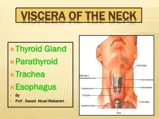
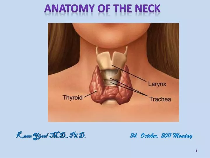
Theneck is thetransitionalareabetweenthebase of thecraniumsuperiorlyandtheclaviclesinferiorly. • Neck is a passagewayformanyimportantstructuresextendingbetweentheheadandthetrunksuch as thelarynxandthethyroidandparathyroidglands.
Theneck is relativelyslendertoallowtheflexibilitynecessarytopositiontheheadtomaximizetheefficiency of itssensoryorgans (mainlytheeyeballs but alsotheears, mouth, andnose). • Thusmanyimportantstructuresarecrowdedtogether in theneck, such as muscles, glands, arteries, veins, nerves, lymphatics, trachea, esophagus, andvertebrae. • Consequently, theneck is a well-knownregion of vulnerability.
Themainarterialbloodflowtotheheadandneck (thecarotidarteries) andtheprincipalvenousdrainage (thejugularveins) lieanterolaterally in theneck. • Carotid/jugularbloodvesselsarethemajorstructurescommonlyinjured in penetratingwounds of theneck.
Thebrachialplexuses of nervesoriginate in theneckandpassinferolaterallytoentertheaxillaeandcontinueintoandsupplytheupperlimbs.
Bones of theNeck • Theskeleton of theneck is formedbythecervicalvertebrae, hyoid bone, manubrium of thesternum, andclavicles. • Thesebonesareparts of theaxialskeletonexcepttheclavicles, whicharepart of theappendicularskeleton.
CervicalVertebrae • 7 cervicalvertebrae form thecervicalregion of thevertebralcolumn, whichenclosesthespinalcordandmeninges. • Thecentrallyplacedvertebralbodies • Supportthehead, andtheintervertebral (IV) articulations • especiallythecraniovertebraljoints at itssuperiorend • Providetheflexibilitynecessarytoallowpositioning of thehead.
HyoidBone (Hyoid) Liesin theanteriorpart of theneck at thelevel of the C3 in theanglebetweenthemandibleandthethyroidcartilage. Suspendedbymusclesthatconnect it tothemandible, styloidprocesses, thyroidcartilage, manubrium, andscapulae. Doesnot articulatewithanyother bone.
It is suspendedfromthestyloidprocesses of thetemporalbonesbythestylohyoidligamentsand is firmlyboundtothethyroidcartilage.
The hyoid consists of a body and greater and lesser horns (L. cornua; the lesser horn is the attachment for the sytlohyoid ligament.). Functionally, the hyoid serves as an attachment for anterior neck muscles and a prop to keep the airway open.
Fascia of theNeck Structuresin theneckaresurroundedby a layer of subcutaneoustissue (superficialfascia) andarecompartmentalizedbylayers of deepcervicalfascia. The knowledge of the organization of the cervical fascia is critical in figuring out where the infections of this region may spread.
Superficial cervical fascia Layerof fattyconnectivetissuethatliesbetweenthedermis of the skin andtheinvestinglayer of deepcervicalfascia. Thistissue is usuallythinnerthan in otherregions, especiallyanteriorly. Containscutaneousnerves, bloodandlymphaticvessels, superficiallymphnodes, andvariableamounts of fat. Anterolaterally, it containstheplatysma.
Theplatysma (G. flatplate) is a broad, thinsheet of muscle in thesubcutaneoustissue of theneck. Theexternaljugularvein (EJV) andthe main cutaneousnerves of theneckaredeeptotheplatysma. Theplatysmacoverstheanterolateralaspect of theneck.
Actingfromitssuperiorattachment, theplatysmatensesthe skin, producingvertical skin ridgesandreleasingpressure on thesuperficialveins. Actingfromitsinferiorattachment, theplatysmahelpsdepressthemandibleanddrawthecorners of themouthinferiorly.
As a muscle of facialexpression, theplatysmaservestoconveytensionorstress. Theplatysma is suppliedbythecervicalbranch of CN VII.
Deepcervicalfascia Thedeepcervicalfasciaconsists of threefasciallayers (sheaths): investing, pretracheal, andprevertebrallayerssupporttheviscera (e.g., thethyroidgland), muscles, vessels, anddeeplymphnodes. Thedeepcervicalfasciaalsocondensesaroundthecommoncarotidarteries, internaljugularveins (IJVs), andvagusnervesto form thecarotidsheath.
Investinglayer of deepcervicalfascia Mostsuperficialdeepfasciallayer, Surroundstheentireneckdeeptothe skin andsubcutaneoustissue. Anteriorly, surroundstheinfrahyoidmuscles.
Theinvestingfascia is attached: • Superiorlytotheexternaloccipitalprotuberanceandthesuperiornuchalline; • Laterallytothemastoidprocessandzygomaticarch; and • Inferiorlytothespine of thescapula, theacromion, theclavicle, andthemanubrium of sternum.
Pretracheallayer of deepcervicalfascia Extendsbetween the hyoid bone and thorax. At the thorax blends with the fibrous pericardium. Has twolayers: Muscularlayerenclosestheinfrahyoidmuscles Viscerallayerencloses the thyroid gland, trachea and esophagus.
Posteriorly, thepretracheallayer is referredto as thebuccopharyngealfascia(a fascia enclosing the pharynx, superior continuation of the fascia covering the buccinator muscle) andseparatesthepharynxandtheesophagusfromtheprevertebrallayer.
Thebuccopharyngealfasciabeginssuperiorly at thebase of theskullandendsinferiorly in thethoraciccavity. Laterally, it blends with the carotid sheath.
Prevertebrallayer of deepcervicalfascia Forms a tubularsheathforthevertebralcolumnandthemusclesassociatedwith it. Extendsbetweenthebase of theskulland T3 vertebra. Attached posteriorlyalongthelength of theligamentumnuchae, andsuperiorlyforms a continuouscircularlineattachingtothebase of theskull.
The prevertebral layer of deep fascia is fixed to the cranial base superiorly. Infero-laterally it extends to axilla and continuous with the axillary sheath enclosing the axillary vessels and the brachial plexus.
CarotidSheath • Tubularfascialinvestment;extendsfromthecranialbasetotheroot of theneck. • Contains : • Commonandinternalcarotidarteries. • Internaljugularvein. • Vagusnerve (CN X). • Somedeepcervicallymphnodes. • Carotidsinusnerve. • Sympatheticnervefibers (carotidperiarterialplexuses)
Thecarotidsheathandpretrachealfasciacommunicatefreelywiththemediastinum of thethoraxinferiorlyandthecranialcavitysuperiorly. • Thesecommunicationsrepresentpotentialpathwaysforthe spread of infectionandextravasatedblood.
Fascial spaces Between the fascial layers in the neck are spaces that may provide a conduit for the spread of infections from the neck to the mediastinum. Three spaces could be involved in this process: Pretrachealspace between the investing layer of cervical fascia and the pretrachealfascia,which passes between the neck and the anterior part of the superior mediastinum; Retropharyngealspace Withinthe prevertebral layer covering the anterior surface of the transverse processes and bodies of the cervical vertebrae.
Retropharyngealspace Largestandmostimportantinterfascialspace in theneck. Potentialspacethatconsists of looseconnectivetissuebetweenthevisceralpart of theprevertebrallayer of deepcervicalfasciaandthebuccopharyngealfasciasurroundingthepharynxsuperficially.
Itpermitsthemovement of pharynx, larynxandesophagus • Theretropharyngealspace is closedwiththeskullbasesuperiorlyandthecarotidsheathlaterally. • Itextendsfromthebase of theskulltotheupperpart of theposteriormediastinum.
CervicalRegions Toallowclearcommunicationregardingthelocation of structures, injuries, orpathologies, theneck is dividedintoregions. Betweenthecranium (mandibleanteriorlyandoccipital bone posteriorly) andtheclavicles, theneck is dividedinto4 majorregions. Basedon theusuallyvisibleand/orpalpableborders of thelargeandrelativelysuperficialSCMandtrapeziusmuscles.
SternocleidomastoidRegion • Thesternocleidomastoid (SCM) muscle is a keymuscularlandmark in theneck, formingthesternocleidomastoidregion [1]. • TheSCM visiblydivideseachside of theneckintotheanterior [2] andlateralcervical[3] regions(anteriorandposteriortriangles). 2 1 3
The SCM is a broad, strap-likemusclethat has twoheads: Sternalheadattachestothemanubrium Clavicularheadattachestotheclavicle
TheSCMsproducemovement at thecraniovertebraljoints, thecervicalintervertebraljoints, or at both. Thecranialattachments of theSCMslieposteriortotheaxis of theatlanto-occipital (AO) joints. Bilateralcontraction of theSCMswillcauseextension of thehead at the AO joints, elevatingthechin Actingbilaterally, theSCMs can alsoflextheneck.
PosteriorCervicalRegion • Theregionposteriortotheanteriorborders of (i.e., correspondingtothearea of) thetrapezius is theposteriorcervicalregion. • Thesuboccipitalregion is deeptothesuperiorpart of thisregion. • Thetrapezius is a large, flattriangularmusclethatcoverstheposterolateralaspect of theneckandthorax.
Thetrapezius is a: • Superficialmuscle of theback • Posterioraxioappendicularmuscle, thatacts on thepectoralgirdle • Cervicalmuscle, that can producemovement of thecranium. • Thetrapeziusattachesthepectoralgirdletothecraniumandthevertebralcolumnandassists in suspending it.
LateralCervicalRegion • Thelateralcervicalregion (posteriortriangle) is bounded: • Anteriorlybytheposteriorborder of the SCM. • Posteriorlybytheanteriorborder of thetrapezius. • Inferiorlybythemiddlethird of theclaviclebetweenthetrapeziusandthe SCM.
Muscles in theLateralCervicalRegion Thefloor of thelateralcervicalregion is usuallyformedbytheprevertebralfasciaoverlyingfourmuscles: spleniuscapitis, levatorscapulae, middlescalene(L. scalenusmedius), andposteriorscalene(L. scalenusposterior).
Nerves of theLateralCervicalRegion Thespinalaccessorynerve (CN XI) passesdeeptothe SCM, supplying it beforeenteringthelateralcervicalregion at orinferiortothejunction of thesuperiorandmiddlethirds of theposteriorborder of the SCM.
Theroots of thebrachialplexus (anterior rami of C5-C8 and T1)appearbetweentheanteriorandthemiddlescalenemuscles. • Thefive rami uniteto form thethreetrunks of thebrachialplexus, whichdescendinferolaterallythroughthelateralcervicalregion. • Theplexusthenenterstheaxilla, providinginnervationformost of theupperlimb.
Vesselsin the LateralCervicalRegion Thelateralbranches of thethyrocervicaltrunk Thethirdpart of thesubclavianartery Partof theoccipitalartery
Veins in lateralcervicalregion Theexternaljugularvein (EJV) is formedbytheunion of theposteriordivision of theretromandibularveinwiththeposteriorauricularvein.
TheEJVterminates in anddrainsintothesubclavianvein. • Thesubclavianvein, themajorvenouschanneldrainingtheupperlimbuniteswiththeIJVto form thebrachiocephalicvein.
Lymphnodes in theLateralCervicalRegion • Lymphfromsuperficialtissues in thelateralcervicalregionentersthesuperficialcervicallymphnodes. • Efferentvesselsfromthesenodesdrainintothedeepcervicallymphnodes, which form a chainembedded in thefascia of thecarotidsheath.
AnteriorCervicalRegion • Theanteriorcervicalregion (anteriortriangle) has thefollowing: • An anteriorboundaryformedbythemedianline of theneck. • A posteriorboundaryformedbytheanteriorborder of the SCM. • A superiorboundaryformedbytheinferiorborder of themandible. • An apexlocated at thejugularnotch in themanubrium. • A roofformedbysubcutaneoustissuecontainingtheplatysma. • A floorformedbythepharynx, larynx, andthyroidgland.
Formorepreciselocalization of structures, theanteriorcervicalregion is subdividedintofoursmallertrianglesbythedigastricandomohyoidmuscles: theunpairedsubmentaltriangle (smen)andthreesmallpairedtriangles—submandibular(sm), carotid (car), andmuscular (mus).
Submentaltriangle • Inferiortothechin • A suprahyoidareaboundedinferiorlybythe body of thehyoidandlaterallybytherightandleftanteriorbellies of thedigastricmuscles. • Containsseveralsmallsubmentallymphnodesandsmallveinsthatuniteto form theanteriorjugularvein. Anteriordigastricmuscles (abd). Mylohyoidmuscle (mh)
Submandibulartriangle • A glandularareabetweentheinferiorborder of themandibleandtheanteriorandposteriorbellies of thedigastricmuscle. • anterior belly of digastric(abd) • posterior belly of digastric (pbd) • Submandibularlymphnodes • Hypoglossalnerve (CN XII) • Nervetothemylohyoidmuscle (a branch of CN V3, whichalsosuppliestheanteriorbelly of thedigastric) • Partsof thefacialarteryandvein • Submentalartery (a branch of thefacialartery)
Musculartriangle (omotrachealtriangle) • Boundedbythesuperiorbelly of theomohyoidmuscle, theanteriorborder of the SCM, andthemedianplane of theneck. Containstheinfrahyoidmusclesandviscera (e.g., thethyroidandparathyroidglands).