Download
1 / 23
230 likes | 375 Views
Channeling through a membrane: How does poliovirus enter cells?. Poliovirus Genome Organization. 743. 7370. 7440. Genomic RNA. AAA. VPg. IRES. 3'-UT. P1. P2. P3. Polyprotein. 3AB. 3C. 3D. 1AB. 1C. 1D. 2A. 2B. 2C. 1A. 1B. VP4. VP2. VP3. VP1. Polymerase. Protease.

E N D
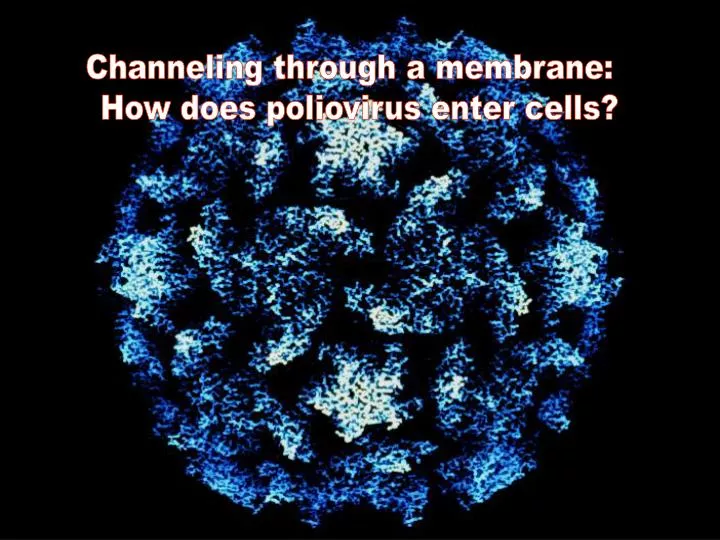
Channeling through a membrane: How does poliovirus enter cells?
Poliovirus Genome Organization 743 7370 7440 Genomic RNA AAA VPg IRES 3'-UT P1 P2 P3 Polyprotein 3AB 3C 3D 1AB 1C 1D 2A 2B 2C 1A 1B VP4 VP2 VP3 VP1 Polymerase Protease Capsid Proteins Replication Proteins
? Attachment (Docking) 160S Particle + PVR RNA release in cytoplasm Infection 160S Native 135S PVR altered 80S Empty Virus entry: The final stages of the morphogenic pathway or "How does a simple nonenveloped virus enter a cell"
“Environmentally Stable” “Environmentally Unstable” “Docking” Entry Active Particle 160S Particle + PVR “Energy” ~100 kcals (Soluble PVR) High Affinity Binding Heat 135S Particle Ea=~120-140 kcals (Kinetically Trapped) Inf+ PVR–
Structural Differences 160S 135S Fricks et al 1990 JV Bubeck et al 2005 JV
“Environmentally Stable” “Environmentally Unstable” 15-20 min ? “Docking” Entry Active Particle 160S Particle Membrane insertion RNA Release + PVR X “Energy” 80S Particle VP4 Mutants 4028T.G 4028T.V 4028T.S 135S Particle
0 30 0 30 min pi WT T.G WT T.S T.V 0 30 0 30 VP1 VP2 VP3 0 30 VP1 VP2 VP3 Western VP4 VP4 Mutant VP4 localize to membranes
Current Distribution @ 40mV Current (pA) Voltage (mV) Voltage Dependence Poliovirus 160S particles form ion channels in lipid bilayers Channel Parameters 1. Conductances 2. Mean open time and probability 3. Voltage dependence (gating, +vs–) 4. Current ion
Control 4028T.S 4028T.G 200ms 1pA 200ms 2 pA Cond. (pS) Mem poten. T1/2-135S Po +40mV 4028T.V WT 15-18 m 0.2 47 + T.G -- -- -- -- 2s 1 pA T.S 15-18 m 0.02 28±10 +/- +40mV T.V 25-30 m ND 15 +/- VP4 entry mutants form ion channels with different properties
“Environmentally Unstable” “Environmentally Stable” 15-20 min “Docking” Entry Active Particle + PVR Ion Channel/ RNA delivery Membrane insertion ? 160S Particle RNA Release “Energy” X 80S Particle VP4 mutants 135S Particle
Phase GFP+ M PVR M PVR Mock Tf PVR/gpi Silver Western Functional reconstitution of purified PVR/gpi onto cells
WT 21°C PVR-GPI +Virus 21°C 5pA 1s PVR-GPI 4028T.G 21°C +Virus 21°C +Virus 2 pA 500ms 31°C Functional Reconstitution of PVR in lipid bilayers
21°C Single Channel Conductance (pS) Temperature (°C) Voltage -Gated Lipid/PVR 21 31 Q10 31°C asolecitin/hPVR No 65±15 ~460 ~7 asolecitin/PVRgpi No 45±7 360±56 8 PE:PS/PVRgpi No 264±5 1450±350 6 Ea (kcals/mole) 160S - 135S 21°C Heat ~120-140 +PVR (sol) ~90-100 +Membrane ~60 Temperature Dependent Conformational Change of PVR - Virus Channel in Lipid Bilayers
Channel model of poliovirus entry 160S 160S PVR PVR 135S delivery of viral RNA genome into cell cytoplasm
Future Directions Define the architecture and structure of the channel 2. Corelate channel behavior in bilayers with mutant entry phenotypes 3. Direct in vivo measurements during infection process
Functional Transitions of Poliovirus Storage and Protection (160S) +PVR ? Delivery (135S) Empty (80S)
Electrophysiology Magdalena Tosteson (Harvard) Khaled Machaca (UAMS) Structure Jim Hogle Dave Filman Doryen Bubeck Mihnea Bostina his collaborators: A. Stevens (NIH) R. McIntosh (UC Boulder) Mutant Analyses Pranav Danthi Xiaojing Ning Qi-Han Li Nicola Moscufo Receptor Yan Huang Hong Wang Anatoli Naumov
Membrane Medium min pi V 0 10 20 30 45 V 0 10 20 30 45 Autorad 100 80 media 60 % Total CPM VP1 VP2 40 VP3 membrane 20 a-VP4 cytoplasm 0 0 10 20 30 40 50 60 VP4 Time Post-Infection (min.) VP4 is inserted into microsomal membranes