Protein Structure Prediction: Computational Analysis & Genomics Study
290 likes | 325 Views
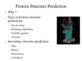
Explore protein structure prediction, secondary to quaternary structure, folding methods, and classification in structural genomics research. Learn about primary amino acid sequences, secondary structure patterns, and tertiary structure formation. Discover various prediction techniques and experimental methods like X-ray crystallography and NMR spectroscopy.

Protein Structure Prediction: Computational Analysis & Genomics Study
E N D
Presentation Transcript
Protein Structure Prediction and Structural Genomics Computer Science Department North Dakota State University Fargo, ND
Outline • Structure of Protein • Prediction Methods • CASP Cup
Protein • Proteins are synthesized as linear chains of amino acids, but they quickly fold into a compact, globular structure. Polypeptide sequence
Each amino acid has two parts, a backbone and a side chain. • The side chain, R, distinguishes the different amino acids. • Backbone is constant for all 20 amino acids. It consists of an amide (--NH2) group, an alpha carbon, and a carboxylic acid (--COOH) group. Peptide bond formation:
Protein Primary structure: • Protein Primary Sequences can be written with a 3-letter code for the 20 amino acids (above) or with a 1-letter code: Ex: Human Insulin A-Chain: GIVEQCCTSICSLYQLENYCN B-Chain: FVNQHLCGSHLVEALYLVCGERGFFYTPKT
Protein Secondary structure • Protein secondary structure refers to regular, repeated patters of folding of the protein backbone. • Patterns result from regular hydrogen bond patterns of backbone atoms.
Protein Secondary Structure • The two most common folding patterns are the alpha helix and the beta sheet.
-helix antiparallel -sheet • Two elements of secondary structure are alpha helices (= =-60o) and beta strands (= -135o,=135o), which associate with other beta strands to form parallel or anti-parallel beta sheets
Secondary Structure • Only two rotatable bonds in protein • The bond between the amide nitrogen and the alpha carbon, referred to as (phi) angle • The bond between the alpha carbon and the carboxyl carbon, referred to as (psi) angle
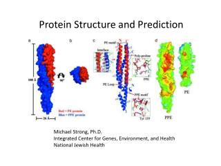
Protein Tertiary Structure • Final shapes of proteins are determined and stabilized by chemical bonds and forces, including weak bonds like Hydrogen bonds, Ionic bonds, Van der Waals bonds, and Hydrophobic attractions. Alpha helices, beta sheets, and turns contribute to the Ribonuclease A tertiary structure. Tertiary Structure of Ribonuclease: A globular protein
Protein Quaternary Structure • The arrangement of the individual subunits of a protein with multiple polypeptide subunits gives the protein a quaternary structure • Ex: Hemoglobin has 2 alpha and 2 beta subunits. • Only proteins with multiple polypeptide subunits can have quaternary structure.
The Goal of Protein Structure Prediction • “The goal of fold assignment and comparative modeling is to assign, using computational methods, each new genome sequence to the known protein fold or structure that it most closely resembles.” • In other words, to class structure into families that share similar folds or motifs and to construct phylogenies.
Significant • Identifying these shared structural motifs can provide significant insight into the functional mechanisms of the protein family. • “The key to understanding the inner workings of cells is to learn the structure of Proteins that form their architecture and carry out their metabolism.” • Comparing proteomics with genomics, it is fair to say that “genes were easy” and the real work of bioinformatics has just begun.
Protein Classification: Families and superfamilies • By definition, proteins that are more than 50% identical in amino acid sequence across their entire length are said to be members of a single family. • Superfamilies are groups of protein families that are related by lower but still detectable levels of sequence similarity (and therefore have a common but more ancient evolutionary origin).
Protein Classification: Folds • Proteins are said to have a common fold if they have the same major secondary structures in the same arrangement and with same topological connections. For example, all alpha proteins, all beta proteins, alpha/beta proteins, membrane and cell surface proteins, etc. • In many respects, the term fold is used synonymously with structural motif but generally refers to larger combinations of secondary structures.
Protein Classification: Enzyme nomenclature • Each enzyme can be assigned a numerical code, such as 3.2.1.14, where the first number specifies the main class, the second and third numbers correspond to specific subclasses, and the final number represents the serial listing of the enzyme in its subclass.
Experimental Techniques • X-ray Crystallography • NMR Spectroscopy • 2D electrophoresis • Mass spectrometry • Protein microarrays
Two Prediction Methods • Protein Folding Model • to simulate the protein folding process at various levels of abstraction which provides insights into the forces that determine protein structure and the folding process. • No algorithm developed to date can determine the native structure of a protein accurately. • Comparative Modeling • sometimes called homology modeling, seeks to predict the structure of a target protein via comparison with the structures of related proteins.
Comparative Modeling Algorithms • DALI (Holm1993) • STRUCTAL (Gerstein1996) • VAST (Gibrat1996) • MINAREA (Falicov1996) • LOCK (Singh1997) • 3dSEARCH (Singh1998)
Prediction Algorithm: 3dSEARCH • Designed to compute fast but approximate alignments of protein structures based on secondary structure elements alone. • The fundamental idea is to represent all secondary structure vectors from all target proteins in a large, highly redundant hash table. Each secondary structure vector from a given query structure can be simultaneously compared to the entire table. • It performed surprisingly well given the simplicity of its technique.
Prediction Algorithm: VAST • Aligning secondary structure elements using graph theory. • Steps of VAST Algorithm • All element pairs (one from each protein) that have the same type are represented as nodes. • Two nodes are connected if the distance and angle within some threshold. • Find the maximal subgraph that are fully connected, which is the pairwise alignment. • Compute alignment score as well as P-value.
Prediction Algorithm: DALI • Attempt to compute the optimal similar contact patterns from a 2-d distance matrices. • Use branch-and-bound algorithm to find an approximate solution.
Prediction Algorithm: STRUCTAL • To minimize the root-mean-square difference (RMSD) between two protein backbones. • Use dynamic programming to minimize.
Prediction Algorithm: MINAREA • To compute a triangulation between the C-a atoms of the two proteins in order to minimize the stretched surface area between their backbones. • Use dynamic programming (DP) to find the minimum.
Prediction Algorithm: LOCK • Attempt to find the optimal rigid-body superposition of two structures such that root-mean-square difference (RMSD) between the aligned C-a atoms is minimized. • An iterative approach that performs a greedy search to the nearest local minimum in alignment space.
Gold Standard for Evaluation • Scope database is being widely used and has been recognized as a current standard in structural classification. (http://pdb.wehi.edu.au/scop) • It has been constructed by visual inspection of all structures in Protein Data Bank (PDB). Four levels, ‘class’, ‘fold’, superfamily’, and ‘family’. ‘Class’ are those that have similar overall secondary structure content.
CASP Competition • CASP competition (Critical Assessment of Techniques for Protein Structure Prediction) http://predictioncenter.llnl.gov/ • Their goal is to help advance the methods of identifying protein structure from sequence.