Download
1 / 85
850 likes | 1.43k Views
Early Concepts: Preformation vs Epigenesis. The question of how a zygote becomes an animal has been asked for centuries.As recently as the 18th century, the prevailing theory was a notion called preformation

E N D
1. Principles of Development Chapter 8
2. Early Concepts: Preformation vs Epigenesis The question of how a zygote becomes an animal has been asked for centuries.
As recently as the 18th century, the prevailing theory was a notion called preformation � the idea that the egg or sperm contains an embryo.
A preformed miniature infant, or �homunculus,� that simply becomes larger during development.
3. Kaspar Friederich Wolff (1759) demonstrated there was no preformed chick in the early egg.
Undifferentiated granular material became arranged into layers.
The layers thickened, thinned, and folded to produce the embryo.
Early Concepts: Preformation vs Epigenesis
4. Epigenesis is the concept that the fertilized egg contains building materials only, somehow assembled by an unknown directing force.
Although current ideas of development are essentially epigenetic in concept, far more is known about what directs growth and differentiation. Early Concepts: Preformation vs Epigenesis
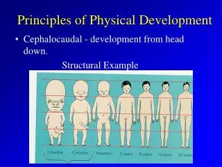
5. Key Events in Development Development describes the changes in an organism from its earliest beginnings through maturity.
Search for commonalities.
6. Key Events in Development Specialization of cell types occurs as a hierarchy of developmental decisions.
Cell types arise from conditions created in preceding stages.
Interactions become increasingly restrictive.
With each new stage:
Each stage limits developmental fate.
Cells lose option to become something different
Said to be determined.
7. Key Events in Development The two basic processes responsible for this progressive subdivision:
Cytoplasmic localization
Induction
8. Fertilization Fertilization is the initial event in development in sexual reproduction.
Union of male and female gametes
Provides for recombination of paternal and maternal genes.
Restores the diploid number.
Activates the egg to begin development.
9. Oocyte Maturation
Egg grows in size by accumulating yolk.
Contains much mRNA, ribosomes, tRNA and elements for protein synthesis.
Morphogenetic determinants direct the activation and repression of specific genes later in post-fertilization development.
Egg nucleus grows in size, bloated with RNA.
Now called the germinal vesicle.
Fertilization
10. Most of these preparations in the egg occur during the prolonged prophase I.
In mammals
Oocyte now has a highly structured system.
After fertilization it will support nutritional requirements of the embryo and direct its development through cleavage.
After meiosis resumes, the egg is ready to fuse its nucleus with the sperm nucleus. Fertilization
11. Fertilization and Activation A century of research has been conducted on marine invertebrates.
Especially sea urchins
12. Contact Between Sperm & Egg Broadcast spawners often release a chemotactic factor that attracts sperm to eggs.
Species specific
Sperm enter the jelly layer.
Egg-recognition proteins on the acrosomal process bind to species-specific sperm receptors on the vitelline envelope.
13. Fertilization in Sea Urchins Prevention of polyspermy � only one sperm can enter.
Fast block
Depolarization of membrane
Slow block
Cortical reaction resulting in fertilization membrane
14. Fertilization in Sea Urchins The cortical reaction follows the fusion of thousands of enzyme-rich cortical granules with the egg membrane.
Cortical granules release contents between the membrane and vitelline envelope.
Creates an osmotic gradient
Water rushes into space
Elevates the envelope
Lifts away all bound sperm except the one sperm that has successfully fused with the egg plasma membrane.
15. Fertilization in Sea Urchins
16. Fertilization in Sea Urchins One cortical granule enzyme causes the vitelline envelope to harden.
Now called the fertilization membrane.
Block to polyspermy is now complete.
Similar process occurs in mammals.
17. Fertilization in Sea Urchins The increased Ca2+ concentration in the egg after the cortical reaction results in an increase in the rates of cellular respiration and protein synthesis.
The egg is activated.
18. Fusion of Pronuclei After sperm and egg membranes fuse, the sperm loses its flagellum.
Enlarged sperm nucleus is the male pronucleus and migrates inward to contact the female pronucleus.
Fusion of male and female pronuclei forms a diploid zygote nucleus.
19. Cleavage Cleavage � rapid cell divisions following fertilization.
Very little growth occurs.
Each cell called a blastomere.
Morula � solid ball of cells. First 5-7 divisions.
20. Polarity The eggs and zygotes of many animals (not mammals) have a definite polarity.
The polarity is defined by the distribution of yolk.
The vegetal pole has the most yolk and the animal pole has the least.
21. Body Axes The development of body axes in frogs is influenced by the polarity of the egg.
22. Amount of Yolk Different types of animals have different amounts of yolk in their eggs.
Isolecithal � very little yolk, even distribution.
Mesolecithal � moderate amount of yolk concentrated at vegetal pole.
Telolecithal � Lots of yolk at vegetal pole.
Centrolecithal � lots of yolk, centrally located.
23. Cleavage in Frogs Cleavage planes usually follow a specific pattern that is relative to the animal and vegetal poles of the zygote.
Animal pole blastomeres are smaller.
Blastocoel in animal hemisphere.
Little yolk, cleavage furrows complete.
Holoblastic cleavage
24. Cleavage in Birds Meroblastic cleavage, incomplete division of the egg.
Occurs in species with yolk-rich eggs, such as reptiles and birds.
Blastoderm � cap of cells on top of yolk.
25. Direct vs. Indirect Development When lots of nourishing yolk is present, embryos develop into a miniature adult.
Direct development
When little yolk is present, young develop into larval stages that can feed.
Indirect development
Mammals have little yolk, but nourish the embryo via the placenta.
26. Blastula A fluid filled cavity, the blastocoel, forms within the embryo � a hollow ball of cells now called a blastula.
27. Gastrulation The morphogenetic process called gastrulation rearranges the cells of a blastula into a three-layered (triploblastic) embryo, called a gastrula, that has a primitive gut.
Diploblastic organisms have two germ layers.
28. Gastrulation The three tissue layers produced by gastrulation are called embryonic germ layers.
The ectoderm forms the outer layer of the gastrula.
Outer surfaces, neural tissue
The endoderm lines the embryonic digestive tract.
The mesoderm partly fills the space between the endoderm and ectoderm.
Muscles, reproductive system
29. Gastrulation � Sea Urchin Gastrulation in a sea urchin produces an embryo with a primitive gut (archenteron) and three germ layers.
Blastopore � open end of gut, becomes anus in deuterostomes.
30. Gastrulation - Frog Result � embryo with gut & 3 germ layers.
More complicated:
Yolk laden cells in vegetal hemisphere.
Blastula wall more than one cell thick.
31. Gastrulation - Chick Gastrulation in the chick is affected by the large amounts of yolk in the egg.
Primitive streak � a groove on the surface along the future anterior-posterior axis.
Functionally equivalent to blastopore lip in frog.
32. Gastrulation - Chick Blastoderm consists of two layers:
Epiblast and hypoblast
Layers separated by a blastocoel
Epiblast forms endoderm and mesoderm.
Cells on surface of embryo form ectoderm.
33. Gastrulation - Mouse In mammals the blastula is called a blastocyst.
Inner cell mass will become the embryo while trophoblast becomes part of the placenta.
Notice that the gastrula is similar to that of the chick.
34. Suites of Developmental Characters Two major groups of triploblastic animals:
Protostomes
Deuterostomes
Differentiated by:
Spiral vs. radial cleavage
Regulative vs. mosaic cleavage
Blastopore becomes mouth vs. anus
Schizocoelous vs. enterocoelous coelom formation.
35. Deuterostome Development Deuterostomes include echinoderms (sea urchins, sea stars etc) and chordates.
Radial cleavage
36. Deuterostome Development Regulative development � the fate of a cell depends on its interactions with neighbors, not what piece of cytoplasm it has. A blastomere isolated early in cleavage is able to from a whole individual.
37. Deuterostome Development Deuterostome means second mouth.
The blastopore becomes the anus and the mouth develops as the second opening.
38. Deuterostome Development The coelom is a body cavity completely surrounded by mesoderm.
Mesoderm & coelom form simultaneously.
In enterocoely, the coelom forms as outpocketing of the gut.
39. Deuterostome Development Typical deuterostomes have coeloms that develop by enterocoely.
Vertebrates use a modified version of schizocoely.
40. Protostome Development Protostomes include flatworms, annelids and molluscs.
Spiral cleavage
41. Protostome Development Mosaic development � cell fate is determined by the components of the cytoplasm found in each blastomere.
Morphogenetic determinants.
An isolated blastomere can�t develop.
42. Protostome Development Protostome means first mouth.
Blastopore becomes the mouth.
The second opening will become the anus.
43. Protostome Development In protostomes, a mesodermal band of tissue forms before the coelom is formed.
The mesoderm splits to form a coelom.
Schizocoely
Not all protostomes have a true coelom.
Pseudocoelomates have a body cavity between mesoderm and endoderm.
Acoelomates have no body cavity at all other than the gut.
44. Two Clades of Protostomes Lophotrochozoan protostomes include annelid worms, molluscs, & some small phyla.
Lophophore � horseshoe shaped feeding structure.
Trochophore larva
Feature all four protostome characteristics.
45. Two Clades of Protostomes The ecdysozoan protostomes include arthropods, roundworms, and other taxa that molt their exoskeletons.
Ecdysis � shedding of the cuticle.
Many do not show spiral cleavage.
46. Building a Body Plan An organism�s development is determined by the genome of the zygote and also by differences that arise between early embryonic cells.
Different genes will be expressed in different cells.
47. Building a Body Plan Uneven distribution of substances in the egg called cytoplasmic determinants results in some of these differences.
Position of cells in the early embryo result in differences as well.
Induction
48. Restriction of Cellular Potency In many species that have cytoplasmic determinants only the zygote is totipotent, capable of developing into all the cell types found in the adult.
49. Restriction of Cellular Potency Unevenly distributed cytoplasmic determinants in the egg cell:
Are important in establishing the body axes.
Set up differences in blastomeres resulting from cleavage.
50. Restriction of Cellular Potency As embryonic development proceeds, the potency of cells becomes progressively more limited in all species.
51. Cell Fate Determination and Pattern Formation by Inductive Signals Once embryonic cell division creates cells that differ from each other,
The cells begin to influence each other�s fates by induction.
52. Induction Induction is the capacity of some cells to cause other cells to develop in a certain way.
Dorsal lip of the blastopore induces neural development.
Primary organizer
53. Spemann-Mangold Experiment Transplanting a piece of dorsal blastopore lip from a salamander gastrula to a ventral or lateral position in another gastrula developed into a notochord & somites and it induced the host ectoderm to form a neural tube.
54. Building a Body Plan Cell differentiation � the specialization of cells in their structure and function.
Morphogenesis � the process by which an animal takes shape and differentiated cells end up in their appropriate locations.
55. Building a Body Plan The sequence includes
Cell movement
Changes in adhesion
Cell proliferation
There is no �hard-wired� master control panel directing development.
Sequence of local patterns in which one step in development is a subunit of another.
Each step in the developmental hierarchy is a necessary preliminary for the next.
56. Hox Genes Hox genes control the subdivision of embryos into regions of different developmental fates along the anteroposterior axis.
Homologous in diverse organisms.
These are master genes that control expression of subordinate genes.
57. Formation of the Vertebrate Limb Inductive signals play a major role in pattern formation � the development of an animal�s spatial organization.
58. Formation of the Vertebrate Limb The molecular cues that control pattern formation, called positional information:
Tell a cell where it is with respect to the animal�s body axes.
Determine how the cell and its descendents respond to future molecular signals.
59. Formation of the Vertebrate Limb The wings and legs of chicks, like all vertebrate limbs begin as bumps of tissue called limb buds.
The embryonic cells within a limb bud respond to positional information indicating location along three axes.
60. Formation of the Vertebrate Limb One limb-bud organizer region is the apical ectodermal ridge (AER).
A thickened area of ectoderm at the tip of the bud.
The second major limb-bud organizer region is the zone of polarizing activity (ZPA).
A block of mesodermal tissue located underneath the ectoderm where the posterior side of the bud is attached to the body.
61. Morphogenesis Morphogenesis is a major aspect of development in both plants and animals but only in animals does it involve the movement of cells.
62. The Cytoskeleton, Cell Motility, and Convergent Extension Changes in the shape of a cell usually involve reorganization of the cytoskeleton.
63. Changes in Cell Shape The formation of the neural tube is affected by microtubules and microfilaments.
64. Cell Migration The cytoskeleton also drives cell migration, or cell crawling.
The active movement of cells from one place to another.
In gastrulation, tissue invagination is caused by changes in both cell shape and cell migration.
65. Evo-Devo Evolutionary developmental biology - evolution is a process in which organisms become different as a result of changes in the genetic control of development.
Genes that control development are similar in diverse groups of animals.
Hox genes
66. Evo-Devo Instead of evolution proceeding by the gradual accumulation of numerous small mutations, could it proceed by relatively few mutations in a few developmental genes?
The induction of legs or eyes by a mutation in one gene suggests that these and other organs can develop as modules.
67. The Common Vertebrate Heritage Vertebrates share a common ancestry and a common pattern of early development.
Vertebrate hallmarks all present briefly.
Dorsal neural tube
Notochord
Pharyngeal gill pouches
Postanal tail
68. Amniotes The embryos of birds, reptiles, and mammals develop within a fluid-filled sac that is contained within a shell or the uterus.
Organisms with these adaptations form a monophyletic group called amniotes.
Allows for embryo to develop away from water.
69. Amniotes In these three types of organisms, the three germ layers also give rise to the four extraembryonic membranes that surround the developing embryo.
70. Amniotes Amnion � fluid filled membranous sac that encloses the embryo. Protects embryo from shock.
Yolk sac � stores yolk and pre-dates the amniotes by millions of years.
71. Amniotes Allantois - storage of metabolic wastes during development.
Chorion - lies beneath the eggshell and encloses the embryo and other extraembryonic membrane.
As embryo grows, the need for oxygen increases.
Allantois and chorion fuse to form a respiratory surface, the chorioallantoic membrane.
Evolution of the shelled amniotic egg made internal fertilization a requirement.
72. The Mammalian Placenta and Early Mammalian Development Most mammalian embryos do not develop within an egg shell.
Develop within the mother�s body.
Most retained in the mother�s body.
Monotremes
Primitive mammals that lay eggs.
Large yolky eggs resembling bird eggs.
Duck-billed platypus and spiny anteater.
73. The Mammalian Placenta and Early Mammalian Development Marsupials
Embryos born at an early stage of development and continue development in abdominal pouch of mother.
Placental Mammals
Represent 94% of the class Mammalia.
Evolution of the placenta required:
Reconstruction of extraembryonic membranes.
Modification of oviduct - expanded region formed a uterus.
74. Mammalian Development The eggs of placental mammals:
Are small and store few nutrients.
Exhibit holoblastic cleavage.
Show no obvious polarity.
75. Mammalian Development Gastrulation and organogenesis resemble the processes in birds and other reptiles.
76. Mammalian Development Early embryonic development in a human proceeds through four stages:
Blastocyst reaches uterus.
Blastocyst implants.
Extraembryonic membranes start to form and gastrulation begins.
Gastrulation has produced a 3-layered embryo.
77. Mammalian Development The extraembryonic membranes in mammals are homologous to those of birds and other reptiles and have similar functions.
78. Mammalian Development Amnion
Surrounds embryo
Secretes fluid in which embryo floats
Yolk sac
Contains no yolk
Source of stem cells that give rise to blood and lymphoid cells
Stem cells migrate to into the developing embryo
Allantois
Not needed to store wastes
Contributes to the formation of the umbilical cord
Chorion
Forms most of the placenta
79. Organogenesis Various regions of the three embryonic germ layers develop into the rudiments of organs during the process of organogenesis.
80. Organogenesis Many different structures are derived from the three embryonic germ layers during organogenesis.
81. Derivatives of Ectoderm: Nervous System and Nerve Growth Just above the notochord (mesoderm), the ectoderm thickens to form a neural plate.
Edges of the neural plate fold up to create an elongated, hollow neural tube.
Anterior end of neural tube enlarges to form the brain and cranial nerves.
Posterior end forms the spinal cord and spinal motor nerves.
82. Derivatives of Ectoderm: Nervous System and Nerve Growth Neural crest cells pinch off from the neural tube.
Give rise to
Portions of cranial nerves
Pigment cells
Cartilage
Bone
Ganglia of the autonomic system
Medulla of the adrenal gland
Parts of other endocrine glands
Neural crest cells are unique to vertebrates.
Important in evolution of the vertebrate head and jaws.
83. Derivatives of Endoderm: Digestive Tube and Survival of Gill Arches During gastrulation, the archenteron forms as the primitive gut.
This endodermal cavity eventually produces:
Digestive tract
Lining of pharynx and lungs
Most of the liver and pancreas
Thyroid, parathyroid glands and thymus
84. Derivatives of Endoderm: Digestive Tube and Survival of Gill Arches Pharyngeal pouches are derivatives of the digestive tract.
Arise in early embryonic development of all vertebrates.
During development, endodermally-lined pharyngeal pouches interact with overlying ectoderm to form gill arches.
In fish, gill arches develop into gills.
In terrestrial vertebrates:
No respiratory function
1st arch and endoderm-lined pouch form upper and lower jaws, and inner ear.
2nd, 3rd, and 4th gill pouches form tonsils, parathyroid gland and thymus.
85. Derivatives of Mesoderm: Support, Movement and the Beating Heart Most muscles arise from mesoderm along each side of the neural tube.
The mesoderm divides into a linear series of somites (38 in humans).
86. Derivatives of Mesoderm: Support, Movement and the Beating Heart The splitting, fusion and migration of somites produce the:
Axial skeleton
Dermis of dorsal skin
Muscles of the back, body wall, and limbs
Heart
Lateral to the somites the mesoderm splits to form the coelom.