Download

1 / 85
880 likes | 1k Views
its a nice and thorough presentation on foot and ankle biomechanics

E N D
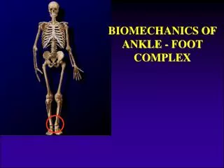
BIOMECHANICS & PATHOMECHANCIS OF ANKLE & FOOT COMPLEX Praveen Shukla MPO-1st Year (2021-22) PDDUNIPPD, New Delhi.
BONES 26 bones & 2 sesamoid bones; divided into 3 functional segments : Forefoot – (Anterior) Metatarsals 5 Phalanges 14 Midfoot – (Middle) Navicular Cuboid Cuneiform 3 Hindfoot – (Posterior) Talus Calcaneus
JOINTS 25 component joints Proximal & distal tibiofibular joints Middle tibio fibular joint- interosseous Ankle (Talo-crural ) joint- HINGED SYNOVIAL JOINT-talus, tibia and fibula(1degree of fredom) Talo-calcaneal (subtalar) joint(inversion/eversion) Talo-navicular joint Calcaneo-cuboid joint 5 tarso-metatarsal joints 5 metatarso-phalangeal joints 9 interphalangeal joints Transverse/Midtarsal joint
Muscles of the ankle : The majority of motion within the foot and ankle is produced by the twelve extrinsic muscles, which originate within the leg and insert within the foot. These muscles are contained within four compartments. The anteriorcompartment consists of four muscles: the tibialis anterior, the extensor digitorumlongus, the extensor hallucislongus, and the peroneustertius. The tibialis anterior and the extensor hallucislongus produce dorsiflexion and inversion of the foot. The peroneustertius produces dorsiflexion and eversionof the foot. The extensor digitorumlongus only produces dorsiflexion of the foot. The lateral compartment is composed of two muscles: the peroneuslongus and the peroneusbrevis, which produce plantarflexion and eversionof the foot. The posterior compartment consists of three muscles: the gastrocnemius, the soleus, and the plantaris, which contribute to plantarflexion of the foot. The deep posterior compartment is composed of three muscles: the tibialis posterior, the flexor digitorumlongus, and the flexor hallucislongus, which produce plantarflexion and inversion of the foot
FUNCTIONS OF ANKLE & FOOT STABILITY Provide stable base of support in variety of weight bearing postures and thus maintain the balance. weight transfer to the ground. MOBILITY Absorbs stress & shock As a loose adapter to accommodate for uneven terrain Act as a rigid lever for forward Progression of body Torque absorber to accommodate for transverse limb rotation PROTECTION– sensation of sole of foot
CARDINAL SYSTEM Dorsiflexion/Planterflexion (horizontal/coronal –axis Sagittal-plane) Abduction/Adduction (vertical axis, transverse plane) Inversion/Eversion (longitudinal/AP- axis, plane- frontal) MOVEMENTS AND AXIS(Ankle Foot Complex)
MOVEMENTS • INVERSION AND EVERSION(subtalar joint movement) Voluntary movements when foot is off the ground. INVERSION- supination+ adduction + some degree of PF- 10 to 30 degrees EVERSION- pronation + abduction + dorsiflexion-10 to 20 degrees
MOVEMENTS PRONATION/SUPINATION: COMPOSITE MOTIONS THAT HAVE COMPONENTS OF CARDINAL MOTIONS AND OCCUR AROUND AN AXIS THAT LIES AT AN ANGLE TO EACH OF THE CARDINAL AXIS. PRONATION: EVERSION+ABDUCTION+DF SUPINATION: INVERSION+ADDUCTION+PF Valgus and varus: terms sometimes used to refer to fixed deformities in the ankle/foot complex Or Movement of the distal segment away form the midline Or Increase in the medial angle between the two reference line-valgus whereas an increase-varus (calcaneovalgus/varus)
Long axis of the foot • A line extending from the middle of the calcaneus to a point between the second and third toes. 1st metatarsal head support two times the weight of each of the other heads.
ANALYSIS OF MOVEMENTS INVERSION : Adduction of calcaneum and navicular with forefoot on the talus +navicular rotation on the head of the talus + gliding of cuboid downwards on the calcaneum+ slight pf of foot EVERSION:( OPPOSITE MOVEMENTS)- Abduction of calcaneum and navicular with forefoot on the talus + navicular rotation and gliding of cuboid upward on the calaneum+ slight df of the foot
Under normal and upright standing conditions the axis of the tibio-talar articulations is perfectly horizontal and oblique as there is normal torsion or twist in the distal tibia in relation to the tibia’s proximal plateau. The knee joint is also horizontal, but the two axis are not parallel. Due to this tibial torsion the foot is outward rotated in relation to the knee joint. Because of this obliquity of the ankle joint axis in upright standing position, the foot moves at the ankle joint upward and medially in dorsiflexion and downward and laterally in planter flexion.
ANKLE, KNEE AND HIP TORSION Axis of motion of hip ,knee and ankle are not parallel but are in torsion to each other if seen from the top. Femoral torsion=12 degree Tibial torsion= 23 degree (from the knee axis)
MEDIAL LONGITUDINAL ARCH • HIGHER ,MORE MOBILE AND RESILIENT THAN LATERAL ARCH • CALCANEUM-TALUS-NAVICULAR- 3 CUNIFORMS-INNER THREE METATARSALS
Truss and tie rod structure • The foot can be considered to function as a truss and tie-rod, with the calcaneus and talus serving as the posterior strut, the remainder to the tarsals and MT forms the ant. Strut while the plantar aponeurosis serving as a tensed tie-rod. Weighting the foot will compress the struts and create additional tension in the tie-rod.
LATERAL LONGITUDINAL ARCH LOW IN HIGHT LIMITED MOBILITY CALCANEUM-CUBOID-OUTER TWO METATARSALS(4,5th)
TRANSVERSE ARCHES • SIDE TO SIDE CONCAVITY • POSTERIOR TRANSVERSE ARCH-formed by the tarsals and posterior part of metatarsals-incomplete half dome(completed when both the feet are joined together • ANTERIOR TRANSVERSE ARCH- formed by the heads of the five metatarsals-complete
FUNCIONS OF ARCHES PROVIDE A PLIABLE PLATEFORM TO SUPPORT THE BODY WIGHT FORWARD PROPULSION OF BODY-walking and running SHOCK ABSORPTION-jumping PROPER WIGHT DISTRIBUTION- heel,balls of the toes Concavity of the arches protects the soft tissues, nerves and blood vessels in the sole THEY ARE BASICALLY FORMED AND MAINTAINED BY THE BONES, LIGAMENTS, MUSCLES, TENDONS AND APPONEUROSIS.
ANKLE JOINT (Talocrural Joint) • Formed between the distal tibia and fibula proximally and the body of the talus distally. 90-95% forces passes through talus and tibia while 5-10% passes through talus and fibula • A synovial hinge joint with a joint capsule and associated ligaments • Single oblique axis with one degree of freedom • Planter flexion/dorsiflexion
Dorsiflexion: 10-20 degree Planter flexion:20-50 degree close packed position of the ankle joint- dorsiflexion, most stable ,withstand compressive forces up to 450% of the body weight. Loose packed position: planter flexion, less stable(high incidence of ankle sprain) Pf/df are limited to soft tissue restrictions , e.g. active or passive tension in the gastroc and soleus limits the d.f. whereas tension in the tibialis anterior , extensor halluicislongus and ext. digitorumlongus limit the p.f. Df is more limited with the knee in extension than with the knee in flexion because the gastorc muscle is lengthened over two joints when the knee is extended. ANKLE JOINT FUNCTION
SUBTALAR JOINT(talocalcaneal joint) • ST Joint axis runs obliquely from dorsum of the foot to planter surface of heel, forming an angle of 45 degree from the transverse plane and 16 degree from the long axis of the foot(sagittal plane) • Has 3 articulations(anterior, posterior-largest and medial)
OPEN CHAIN KINETIC MOTION non weight bearing: triplanar motion. 1.Pronation-calcaneus eversion(frontal plane), abduction(transverse plane)and dorsiflexion (sagittal plane) 2. Supination- reverse motion viz. calcaneal inversion, adduction and pf of calcaneus and foot
CLOSED CHAIN KINETIC MOTION Foot and heel are in contact with the ground(weight bearing) Pronation- calcaneus eversion, adduction and planter flexion(talus head), associated with internal rotation of tibia, abduction and inversion of the forefoot on the hindfoot and depression of the ML arch Supination- reverse- calcaneal inversion, abduction and dorsiflexion (talus head)with abduction and eversion of the forefoot on the hiddoot and elevation of the ML arch
SUMMARY OF STJ MOTION Non Weight bearing Weight bearing Supination : calcaneal inversion(varus) calcaneal inversion(varus) calcaneal adduction Talar abduction(lateral rotation) calcanealplanterflexionTalardorsiflexion Tibiofibular lateral rotation Pronation: calcanealeversion(valgus) calcanealeversion(valgus) calcaneal abduction talar adduction(medial rotation) calcanealdorsiflexiontalarplanterflextion tibiofibular medial rotation
STJ- MOVEMENTS • These subtalar component motions are coupled and cannot occur independently and must occur simultaneously • The contribution of each of the coupled movements to supination or pronation will depend greatly on individual differences in inclination of the subtalar axis. • Higher inclination of STJ axixw.r.t. horizontal will result in decrease ROM. • (Cavus-rigid Vs Flat Foot-mobile)
SUBTALAR NEUTRAL • When posterior calcaneus and posterior midline of the leg coincide each other. ( 0 degree)- midstance • 5-10 degrees calcanealeversion(valgus) and 20-30 degrees calcaneal inversion(varus)- individuals without impairment • Variability in the inclination of the subtalar axis determines the range of the coupled components of subtalar motion.
STJ SHOULD BE IN NEUTRAL POSITION DURING MIDSTANCE FACTORS INFLUENCES STJ POSITION: 1. STRUCTUAL DEVAITON(FEMORAL OR TIBIAL ROTATION) . EXTRINSIC FACTRORS LIKE FOOTWEAR, RUNNING SURFACE, ACTIVITY LEVEL . PHYSIOLOGICAL FACTORS SUCH AS OBESITY OR DEISEASE. ST NEUTRAL
MIDTARSAL/TRANSVERSE TARSAL JOINT • FORMED BY –TALONAVICULAR AND CALCANEOCUBOID JOINTS (S-shaped line) • Talonavicular joint-posterior concave surface of navicular and anterior convex head of talus(ball and socket) • Calcaneocuboid joint-formed by the anterior calcaneus and posterior cuboid bone
MIDTARSAL/TRANSVERSE TARSAL JOINT-Axes Longitudinal axis-15 degree inclined from the transverse plane and 9 degree medially from the sagittal plane Triplanermovments-supination/pronation and coupled component movements