Download
1 / 68
680 likes | 888 Views
Melanoma Overview 2009. Frances Collichio Associate Professor, University of North Carolina, Chapel Hill Disclosure: OncoVex Novartis. What is Melanoma?. A Cancer of Melanocytes All Melanomas are malignant Melanocytes? Cells that Make Pigment

E N D
Melanoma Overview 2009 Frances Collichio Associate Professor, University of North Carolina, Chapel Hill Disclosure: OncoVex Novartis
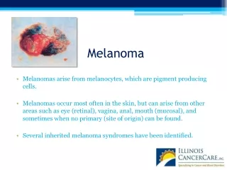
What is Melanoma? • A Cancer of Melanocytes • All Melanomas are malignant • Melanocytes? • Cells that Make Pigment • Melanoma therefore can start from any pigmented cell.
Extracutaneous Melanomas, • Mucosal • GI tract, Head and Neck, vagina • Older pts • Poorer px • Disporportionally non-white
Ocular • Uveal • Ciliary body/Choroidal • Poor prognosis • Iris • Better px
Unknown Primary • 2-4% of all melanoma • 9% of melanoma with lymph node involvement • Search for the primary • Ocular exam when there are liver mets When you cannot find the primary, treat these as if they started in the skin.
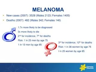
“Silent”NationalEpidemic • The incidence per year is rising faster than any other cancer! Rate/100,000 1:1500 1:600 1:250 1:150 1:135 1:75 1:65 1:60 estimated Lifetime Risk
2007 Estimated US Cancer Cases* Men766,860 Women678,060 • 26% Breast • 15% Lung & bronchus • 11% Colon & rectum • 6% Uterine corpus • 4% Non-Hodgkin lymphoma • 4% Melanoma of skin • 4% Thyroid • 3% Ovary • 3% Kidney • 3% Leukemia • 21% All Other Sites Prostate 29% Lung & bronchus 15% Colon & rectum 10% Urinary bladder 7% Non-Hodgkin 4% lymphoma Melanoma of skin 4% Kidney 4% Leukemia 3% Oral cavity 3% Pancreas 2% All Other Sites 19% *Excludes basal and squamous cell skin cancers and in situ carcinomas except urinary bladder. Source: American Cancer Society, 2007.
Melanoma US 2008 • 108,000 estimated new cases • 62,480 invasive • 48,290 in situ • 8,420 deaths www.cancer.org
Etiology • Inheritance -Chromosome 9 (10-40%) • Environment • Sun • Greater than 3 blistering sunburns under age 20 • Freckles on the back • Tanning Booths • Genes-Environment Interactions
Melanoma A—Asymmetry B—Border C—Color D—Diameter E—Evolution: A changing mole
Nodular Amelanotic Melanoma Invasive Melanoma
After diagnosing a skin lesion as melanoma, what is next? • Surgery on the primary by an experience surgeon or dermatologist • The margins of resection around the primary depend on the depth of the primary • Less than 1mm deep, 1cm margin • 1 to 2mm deep, a 1 to 2cm margin • Greater than 2 mm deep, a 2 cm margin
Staging, 2002 AJCC • T • Breslow depth • Clark level---used only in T1 melanomas • Ulceration • N • N1: Metastasis to one node • N2: Metastasis to two or three nodes • N3: Mets to 4 or more, or matted nodes, or in transit disease, or satellites (tumor w/in 5cm of the primary). • Micromets are defined by sentinel node. Macro mets are clinically detectable. • M • M1a-skin, subcutaneous tissue or distant lymph nodes • M1b-lung • M1c-other sites
T stage • TX: Primary cannot be assessed (shave bx) • T0: no evidence of primary • T1 1mm or less • T1a: 1mm or less and Clarks level II or III w/o ulceration • T2 >1mm and <2mm • T3 >2mm and <4mm • T4 >4mm • A:w/o ulceration • B:w ulceration
Nodal Stage • N • N1: Metastasis to one node • N1a:micromet • N1b: Macromet • N2: Metastasis to two or three nodes • N2a: Micromet • N2b: Macromet • N2c:in-transit met(s)/satellite(s) without metastatic lymph nodes • N3: Mets to 4 or more, or matted nodes, or in transit disease, or satellites (tumor w/in 5cm of the primary).
Sentinel Node procedure • Stages the patient • Directs further diagnostic studies • May have a therapeutic impact • Avoid unnecessarily complex dissections.
Sentinel Lymph Node Evaluation Lymph node serially sectioned while fresh Alternate sections paraffin-embedded; remainder snap frozen MS01-1234 Level 1 S-100 Doe, John MS01-1234 Level 2 H&E Doe, John MS01-1234 Level 3 H&E Doe, John MS01-1234 Level 4 H&E Doe, John MS01-1234 Level 5 H&E Doe, John MS01-1234 Level 6 H&E Doe, John MS01-1234 Level 7 S-100 Doe, John Five H&E-stained sections flanked by two S-100 sections
Indications for the Sentinel Lymph Node Procedure • Thin melanomas (< 1 mm) • Consider for ulcerated primaries or Clarks IV, V • Intermediate thickness melanomas (1-4 mm) • Risk: 18-20% • Thick melanomas (>4 mm) • Risk: 40%+ nodal, 15-20% systemic
Patients with Postive Lymph Nodes • Have completion Lymph node surgery.
After treating the primary and completing lymph node surgery, what is next?
Now • You know the T stage • You know the N stage • You base the extent of additional studies on those two facts.
Additional Studies? • Stage I to IIA: (up to a 4mm thick with no ulceration) No additional Studies • Stage IIB, IIC: Additional Studies as clinically indicated (CT, PET, MRI) • Stage III: Baseline studies for staging or symptoms---Chest x-ray, CT + PET, MRI brain
Why PET/CT • CT commonly used alone • Can miss visceral/lung metastases Reinhardt, MJ et al. J Clin Oncol 2006;24:1178
Treatment of Stage IIb (T4), IIc, and III Melanoma when all of the visible tumor has been removed • Observation • Clinical Trial • Or Interferon alpha
Adjuvant Therapy • Only one FDA Approved Therapy • Interferon-a 2b (Brand Name: Intron) • Meta-Analysis ~ 10% Absolute Benefit in RFS • Improved QoL versus Observation • Cost Effective • Patient Preference compared to increased risk • 3% survival benefit but not statistically significant Cole, BF et al. J Clin Oncol 1996;14:2666 Hillner, BE et al. J Clin Oncol 1997;15:2351 Killbridge, KL et al. J Clin Oncol 2001;19:812
Interferon Regimen IFNa 2b 20 Million Units/m2/Day IV (15MU often new starting point) 5 Days per week for 4 weeks 10 Million Units/m2/Day Subcutaneous 3 times per week for 11 months Side Effects: Flu-like Sx, Hepatic Transaminitis, Fatigue, HA, Nausea, Wt Loss, Myelosuppression, Depression
Adjuvant Radiation Therapy • Consider RT to the nodal basin when there are multiple nodes involved or extranodal extension (category IIb evidence NCCN)
Stage IV • Median survival is 9 months and less than 5% probability of survival beyond 5 years.
Stage IV disease local Therapies • Surgery • Best for skin/lymph nodes>lung>GI tract • Radiation • Palliation of symptomatic sites • High dose fractions (?) • Brain Mets
Systemic Treatments • Where is this going • Genetic Footprint • What Categories of treatment do we see? • Immune-therapy sensitive • Chemotherapy • Targetted Therapy
MAPK and PI3K/AKT pathways Cell Membrane--------------------------------------------------------- SHC PI3K→AKT RAS PTEN ↓ BRAF MEK ½ CCND1 MAPK3 ↓ MAPK1 CDK4/6 ↓ ↓ ↓ VEG CFOS ELK Proliferation Proliferation ↓ Cell cycle progression
MAPK and PI3K/AKT pathways • Ras/Ras PI3/AKT Cutaneous Not chronically sun exposed Cutaneous, Chronically sun exposed Mucosal, Acral Proliferative pathway Antiapototic pathway CDKN2A gene mutations CDK4 amplifcation in acral and mucosal
KIT • Receptor tyrosine kinase • Critical regulator of growth of melanocytes. • Dysfunctional KIT pigmentary defects. • Expression is lost in nevi • Amplification in chronically sun exposed melanoma • Amplification in KIT exons 11, 13, 17 and 18 may correlate with Rx response
KIT Case • 79 yo rectal melanoma • 12/06 recurrence, anal rectal junction and near the kidney • Strong staining for KIT • KIT exons 11, 13, 17 were amplified • Additional peak in exon 11 • Imatinib 400mg daily • Marked improvement in all disease J Clin Oncol 2008
Imatinib Study • For patients with KIT amplification or mutation • Gleevac 400mg PO daily
Immunotherapy • IL2 • Vaccines • Other Modulators of the immune system • CTLA 4 • New agents
Immune Modulating AgentsHigh Dose IL 2 • Increases CD 4 positive cells • Pooled analysis ORR 14% • CR 5% • Highly selected patients • Most durable response of the known therapies • Toxic. Requires ICU care and expert personnel
Any hope for Vaccines ? • Stage IIIB and IV • OncoVEXGM-CSF will be administered by injection into all injectable cutaneous, subcutaneous or nodal lesions lesions, every two weeks. • This vaccine uses Herpes virus proteins as a co-stimulant