Download
1 / 54
540 likes | 655 Views
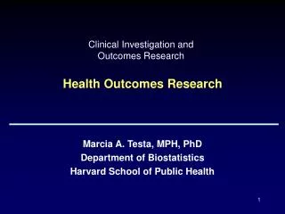
Clinical Investigation and Outcomes Research Analysis of Physiologic and Pharmacologic Data. Marcia A. Testa, MPH, PhD Department of Biostatistics Harvard School of Public Health. Objective of Presentation.

E N D
Clinical Investigation and Outcomes ResearchAnalysis of Physiologic and Pharmacologic Data Marcia A. Testa, MPH, PhD Department of Biostatistics Harvard School of Public Health
Objective of Presentation • Introduce analytical methods for the special case where biomedical data are collected during a session which contains: • repeated observations over time • numerous, frequently sampled data points • measures collected over a relatively short interval of time (several hours or days) within one session • commonly, more measures per session per subject, than subjects overall
Intensively Sampled Data • Data collected during a physiology, monitoring or pharmacologic study over several hours or days with measurement every 1, 5, 10, 15, 30 or 60 minutes, or as a continuous function • Each session may be repeated at weekly or monthly intervals to investigate the effects of interventions as part of clinical trials or treatment assessment, and to correlate session summary parameters with clinical events, morbidity and mortality • In physiologic research, these data are often referred to as “complex physiologic signals”
ECG BP Why Study Signals? Physiologic signals and time series reveal aspects of health, disease, biotoxicity and aging not captured by static measures. Time = 2 seconds Raw (original) signals are of interest as means of • developing new biomarkers • measuring parameters of known interest • developing new insights into basic • mechanisms of human physiology
Periodic Functions Physiologic Response Time (minutes) Response may represent a periodic function such as this graph of interstride intervals for a patient with Huntington’s disease, or a smooth function in response to a stimulus such as oral drug administration.
Smooth Functions Ka = Absorption Constant Ke = Elimination Constant Ka/Ke=10 Ka/Ke=1 Ka/Ke=0.1 Plasma concentration Ka/Ke=0.01 Oral Drug
Intensive Data: Cardiology Studies • Continuous recording: ECG is recorded continuously during the entire testing period. • Event monitor, or loop recording: ECG is recorded only when the patient starts the recording, when symptoms are felt.
A Complex Signal Dataset Physiologic time series, such as this series of cardiac interbeat (RR) intervals measured over 24 hours, can capture some of the information lost in summary statistics. Data from the NHLBI Cardiac Arrhythmia Suppression Trial (CAST) RR Interval Sub-study Database
Example 1: Heart Rate Dynamics Pathology can affect physiologic recordings in unexpected and interesting ways. Analysis of complex signals can extract information hidden in data. Figure shows shows the instantaneous heart rates of four subjects. The plot of heart rate (beats/min) versus time (min) is called a tachogram. Of the four tachograms shown, only one signal is from a healthy person. Can you tell which it is?
In A and C we can see a rather periodic signal, with low variability of its values. In case C, there is a pattern of periodic oscillations (1/min), which is associated with Cheyne-Stokes breathing. The healthy record B is characterized by a rather rough and ‘patchy’ configuration, attributed to fractal properties of the heart rate signal. The breakdown of such behavior (fractal dynamics) can lead to either excessive regularity (A &C) or uncorrelated randomness (D). Excessive regularity Healthy heart rate Excessive regularity Uncorrelated Randomness
Example 2: Ambulatory ECG Schedule of study events is shown in panel A. Panel B shows in-hospital activity schedules on the two activity days. AEM indicates ambulatory ECG monitoring. Vertical arrows represent timing of venous sampling. A B
Regular Activity Day Example 2: Rates of Ambulatory Ischemia – Bar Graphs and Polynomial Regression Parker JD, Testa MA, Jimenez AH, Tofler GH, Muller JE, Parker JO and Stone PH. Morning increase in ambulatory ischemia in patients with stable coronary artery disease: Importance of physical activity and increased cardiac demand. Circulation 1994;89:604-614.
Example 2: Rates of Ambulatory Ischemia – Bar Graphs and Polynomial Regression Delayed Activity Day Parker JD, Testa MA, Jimenez AH, Tofler GH, Muller JE, Parker JO and Stone PH. Morning increase in ambulatory ischemia in patients with stable coronary artery disease: Importance of physical activity and increased cardiac demand. Circulation 1994;89:604-614.
Regular Activity Day A Example 2: Ambulatory ECG Bar graphs show frequency of episodes of ambulatory ischemia during therapy with placebo and nadolol on the two activity days. Panel A, Regular activity day;panel B, delayed activity day. Delayed Activity Day B
Example 2: Minute by Minute Heart Rate Placebo Nadolol Parker JD, Testa MA, Jimenez AH, Tofler GH, Muller JE, Parker JO and Stone PH. Morning increase in ambulatory ischemia in patients with stable coronary artery disease: Importance of physical activity and increased cardiac demand. Circulation 1994;89:604-614.
Example 2: Minute by Minute Heart Rate Placebo Nadolol Parker JD, Testa MA, Jimenez AH, Tofler GH, Muller JE, Parker JO and Stone PH. Morning increase in ambulatory ischemia in patients with stable coronary artery disease: Importance of physical activity and increased cardiac demand. Circulation 1994;89:604-614.
Example 3: Continuous Glucose Monitoring in Diabetes Continuing Glucose Monitoring Systems Each colored line represents 5-minute glucose samples for a different day of the week.
Intensively sampled data can arise from many sources during the same clinical study Continuing Glucose Monitoring Systems Glucose Meter E-Diary
Example 4: Pharmacokinetics • Pharmacokinetics provides good general framework for the family of models which involves extracting parameters representative of biological processes • Drug absorption, distribution, metabolism and excretion • Intensity and duration of therapeutic and toxic effects of many drugs are closely related to their biological availability and disposition
Elimination phase Absorption phase Example 4: Plasma Concentration of Drug after Oral Administration
Steps in Analysis • Collect raw signal data (e.g., heart rate, glucose, plasma concentration) and transfer to relational database for estimation of parameters • Estimate signal parameters (e.g., heart rate variability, glucose variability, pharmacokinetic rate constants) using analytical programs • Use estimated parameters as dependent measures for prediction of health outcome or mortality (Exposed vs Unexposed), or determine how treatment (e.g., beta blocker) changes signal and how that change impacts health outcome, clinical event or mortality (Experimental vs. Control)
Estimating HRV Parameters Hear Rate Variability (HRV) Adapted from Goldberger AL. Fractals dynamics in phy-siology: Alterations with disease and aging. PNAS 2002; 99: 2466-2472, downloaded from www.physionet.org.
HRV: Time-Domain Methods • Based upon beat-to-beat or RR intervals • SDRR: standard deviation (SD) of RR intervals over 24 hours • SDARR: SD of average RR intervals calculated over short periods ( 5 mins) • RR50: number of pairs of successive RRs that differ by more than 50 minutes.
HRV: Frequency-Domain Methods • Fast Fourier transform • High Frequency band (HF) between 0.15 and 0.4 Hz. HF is driven by respiration and appears to derive mainly from vagal activity (parasympathetic nervous system). • Low Frequency band (LF) between 0.04 and 0.15 Hz. LF derives from both parasympathetic and sympathetic activity and has been hypothesized to reflect the delay in the baroreceptor loop.
HRV: Frequency-Domain Parameters • Fast Fourier transform • Very Low Frequency band (VLF) band between 0.0033 and 0.04 Hz. The origin of VLF is not well known. • Ultra Low Frequency (ULF) band between 0 and 0.0033 Hz. The major background of ULF is day–night variation and therefore is only expressed in 24-hour recordings. • The ratio of low-to-high frequency spectra power(LF/HF) has been proposed as an index of sympathetic to parasympathetic balance of heart rate fluctuation, but this is controversial because of the lack of understanding of the mechanisms for the LF component.
HRV: Non-linear Methods • Poincaré plot. Each data point represents a pair successive beats, the x-axis is the current RR interval, while the y-axis is the previous RR interval. • HRV is quantified by fitting mathematically defined geometric shapes to the data. • Other methods used are the correlation dimension, nonlinear predictability, point wise correlation dimension and approximate entropy.
The abscissa represents the RR interval of the current normal beat and ordinate represents the RR interval of the succeeding normal beat. An ellipse is fitted to the data points and the Poincaré plot indices are calculated by estimating the short diameter (SD1), the long diameter (SD2) and the ratio of the short and long diameters (SD1/SD2 ratio) of the fitted ellipse Poincaré plot
Pharmacokinetic Processes • Liberation – the release of the drug from its dosage form • Absorption – the movement of drug from the site of administration to the blood circulation • Distribution – the process by which the drug diffuses or is transferred from intravascular space to extravascular space (body tissues) • Metabolism – the chemical conversion of drugs into compounds that cab be eliminated • Excretion – the elimination of unchanged drug or metabolite from the body via renal, biliary, or pulmonary processes.
Elimination Constant First order elimination, rate is proportional to concentration. The elimination rate constant Kel represents the portion of the drug eliminated per unit time.
Elimination Constant (Log scale) The slope of the line of the concentration plotted on the log scale correlates with Kel. Kel = ln(Peak/Trough)/time (P-T))
First Order Process T = 0, C = 100 Loss from 1 to 2 is proportional to C dC dt L(2, 1) First order rate constant SIDE A SIDE B COMP 1 COMP 2
How do you estimate parameters? There are several software packages that can be used to estimate parameters – such as those from www.adinstruments.com as shown here.
How do I estimate parameters? There are several software packages that can be used to estimate parameter – such as those from www.adinstruments.com as shown here.
Pharmacokinetic Analysis Software Several different packages may be used. e.g.,(shown) http://www.summitpk.com/
www.physionet.org NIH has a data archive and free software.
What is Physionet? • NIH-sponsored Research (Harvard, BU, McGill) established in 1999 • Freely available physiologic data and open-source software • PhysioBank: 4000 recordings of digitized physiologic signals and time series, over 40 databases • PhysioToolkit: Open source software
Physionet Tutorials and Datahttp://www.physionet.org/tutorials/hrv/
Data for Sample Patient – 4 Days Is the “mean” the best way to summarize these data?
Data for Sample Patient – Session Week 12-- there are many parameters that could be estimated for each subject
Summarize the Raw Data • The individual daily curves should be summarized to obtain signal parameters meaningful to the research objectives • Examples • Mean, Max, Minimum for each day • Percent > 180 mg/dl (hyperglycemia) • Percent < 36 mg/dl (severe hypoglycemia) • Intraday standard deviation (glucose variability) • Area above and below defined thresholds
Simple Numeric Transformations /BREAK=Patient_ID by CGMS_num by Date by Nocturnal /Sensor_Glucose = NU(Sensor_Glucose) /Sensor_1 = MEAN(Sensor_Glucose) /Sensor_2 = MEDIAN(Sensor_Glucose) /Sensor_3 = SD(Sensor_Glucose) /Sensor_4 = MIN(Sensor_Glucose) /Sensor_5 = MAX(Sensor_Glucose) /Sensor_6 = PGT(Sensor_Glucose 140) /Sensor_7 = PLT(Sensor_Glucose 70) /Sensor_8 = PGT(Sensor_Glucose 180) /Sensor_9 = PLT(Sensor_Glucose 60) /Sensor_10 = PLT(Sensor_Glucose 50) /Sensor_11 = PGT(Sensor_Glucose 300) /Sensor_12 = MEAN(Sens_gluHI) /Sensor_13= MEAN(Sens_gluLO) /Sensor_14 = SD(Sens_gluHI) /Sensor_15= SD(Sens_gluLO) Data Reduction from 1000’s to only 15 measures per subject – all representing a different parameter of the CGMS profile curve Code Shown – using functions from a common statistics package or Excel.
More Sophisticated Modeling Techniques:Fourier Series Start with a sine wave: Build a model using Fourier Series The theory of Fourier series lies in the idea that most signals, can be represented as a sum of sine waves
CGM Daily Measures • Mean Glucose (24-hour, day-time, nocturnal) • Mean Glucose Standard Deviation • Mean amplitude glucose excursions (MAGE) • Low blood glucose index (LBGI) • High blood glucose index (HBGI) • AUC of BG < 70 mg/dL (3.9 mmol/L) and < 50 mg/dL (2.8 mmol/L) • Nocturnal hypoglycemia – measures < 36, 50, or 70 mg/dL during late night and early morning (sleep time)
CGM Post-Prandial Measures Some summary parameters may be in response to meals. • Meal Interval Start Glucose • Meal Interval Start Time • Pre-Meal Insulin Dose • Meal Type • Glucose (C0 (mg/dl), Time (0) • Glucose Cmax (mg/dl), Glucose Tmax (min), Glucose (Cmax - C0), Glucose (Tmax - T0), • Glucose Cmin (mg/dl - trough) • Glucose Tmin (min) • Glucose (Cmax – Cmin ) • Glucose Upstroke (Appearance Rate) • Glucose Downstroke ( Elimination Rate)
Data for Sample Patient • The patient had three sessions of continuous glucose monitoring with each session lasting several days. • Below are the overall mean glucoses for each of the sessions Case Week Initials Mid Interval Date Glucose 100000.0 0 XYZ 20-OCT-2009 185.33 100000.0 12 XYZ 15-JAN-2010 133.63 100000.0 24 XYZ 06-APR-20`0 133.90
Graph of Mean Glucose at Weeks 0, 12 and 24 for Patient 100000 0 12 24 Weeks