Download

1 / 44
470 likes | 842 Views
Acquired Heart Disease. Tricia Santos MS3 Diagnostic Radiology December 2005. Diagnosing heart disease the “old fashioned” way. History Physical Exam Chest Radiograph. Approach to evaluation of the Heart on Chest Radiograph. Evaluate the heart for: Pericardial disease

E N D
Acquired Heart Disease Tricia Santos MS3 Diagnostic Radiology December 2005
Diagnosing heart disease the “old fashioned” way History Physical Exam Chest Radiograph
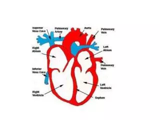
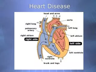
Approach to evaluation of the Heart on Chest Radiograph • Evaluate the heart for: • Pericardial disease • Myocardial disease • Valvular disease • Evaluate the vessels for: • Pressure and flow changes • Intravascular volume status • Edema
Size Matters When looking for heart disease, first ask yourself, “Is the heart big or small?”
Pericardial and Myocardial disease= Global enlargement Small Heart • Constrictive Pericarditis • Restrictive Cardiomyopathy Big Heart • Pericardial Effusion • Myocardial Failure
Small HeartPericardial or Myocardial Disease? • Use physical exam to differentiate • Kussmaul’s sign and pericardial knock are consistent with constrictive pericarditis Pericardial Calcifications Small Heart
Globally Enlarged HeartPericardial Disease • Pericardial Effusion • “Oreo” Sign • Fluid collection between epicardial and retrosternal fat pads • WIDE vascular pedicle • RA pressures are high due to constriction and therefore do not allow blood to easily return to the RA
Globally Enlarged HeartMyocardial Disease • Myocardial Failure • NARROW vascular pedicle • Patients are usually on diuretics • Leads and Lines • Outline the walls of the chambers if no effusion is present
Myocardial Failure or Pericardial Effusion? Wide Vascular Pedicle Visible Borders of Mediastinum Pericardial Effusion
Myocardial Failure or Pericardial Effusion? Gehlbach, Brian K., et al. The Pulmonary Manifestations of Left Heart Failure. Chest. 2004; 125: 669-682.
Myocardial Failure or Pericardial Effusion? Globally enlarged heart Narrow VPW Myocardial Failure Gehlbach, Brian K., et al. The Pulmonary Manifestations of Left Heart Failure. Chest. 2004; 125: 669-682.
Small/Normal Heart • Valvular Stenosis - Chambers are pressure overloaded - Mild dilation of chambers may be seen, but general hypertrophy is not seen on chest radiograph Big Heart • Valvular Insufficiency - Chambers are volume overloaded - Marked dilation of chambers Valvular Disease = Unequal chamber enlargement
Aortic Stenosis • Chest radiograph • Decreased pulmonary blood flow with normal flow distribution • Narrow vascular pedicle • Increased LVP (may have mild LV enlargement) • Post stenotic dilation of aorta • Physical Exam: • Crescendo/decrescendo systolic murmur (may radiate to clavicles, carotid, or “beauty-sash” distribution) • Pulsus parvus et tardus • Diastolic rumble from associated aortic insufficiency
Aortic Stenosis Why narrow vascular pedicle with decreased pulmonary blood flow? • Low LV output → decreased circulating blood volume → decreased venous return and RV output • Increase in circulating atrionatriuretic factor → decreased total blood volume → decreased venous return and RV output
Mitral Stenosis • Chest radiograph • Mild LA dilation • Increased LAP • Pulmonary flow inversion • LUL oligemia occurs in 16% of patients • Possibly secondary to displaced/compressed LUL veins from LA • Narrow vascular pedicle • Physical Exam • Faint diastolic murmur (rumble) • Opening snap • Loud S1
Atrial Septal Defect…Why? LUL Oligemia Narrow VPW Pulmonary Venous HTN LV Dilation
Mitral Insufficiency • Chest Radiograph • Marked dilation of LA • Pulmonary flow inversion • Physical Exam • Holosystolic blowing murmur • Radiates to axilla • No change with inspiration • S1 and S2 may be inaudible or difficult to hear • Systolic apical thrill
Tricuspid Insufficiency • Chest radiograph • Marked dilation of RA • Wide vascular pedicle • Physical Exam • Holosystolic blowing murmur • Increases with inspiration/increased venous return • Elevated JVP with fused CV wave • Side-to-side head bob • Hepatojugular reflux • Hepatomegaly • Puslatile Liver • Ascites • Peripheral Edema
Evaluate the vessels:Pulmonary Blood Flow • Increased with shunt vascularity • Decreased with cephalization • Flow inversion occurs with chronic left heart failure and mitral stenosis
Normal Pulmonary Flow • Pulmonary veins have no valves, therefore they are directly affected by pressures in the LA • In the upright person, flow is greater in the lower lobes according to the West zones • Gravity makes it more difficult for blood to return to the LA from the lower lobe veins, therefore LL vessels are larger
Pulmonary Flow Inversion • Occurs with long-standing elevated LAP • Actual cause of redirection of blood is unknown • One theory suggests: ↑ LAP → basal edema → ↓ basilar compliance → ↓ negative interstitial pressure → vessels unable to stay open → ↓ diameter of vessels → ↑↑ resistance to flow → blood redirected to upper lobes • Others theorize that the cause is organic • Cardiac output is likely decreased in the presence of cephalization and edema • Flow inversion is not reversible with treatment
Pulmonary Flow Left to Right Shunts • ASD, VSD, and PDA originally shunt blood to the right side of the heart and pulmonary circulation • Pulmonary flow INCREASES • Narrow vascular pedicle secondary to decreased systemic flow • Small aorta due to decreased LV output • PE: Listen for the presence of murmurs • ASD: systolic, fixed split S2 • VSD: loud, harsh, holosystolic • PDA: “machine-like” systolic and diastolic
Decreased with Cephalization Larger vessels Small Vessels
Evaluate the Vessels:Pulmonary Pressures • Pulmonary Venous Hypertension • Caused by subacute to chronic impairment of pulmonary venous drainage, i.e. ↑ LAP • Myocardial dysfunction • Mitral valve disease • Obstruction • Secondary signs include septal thickening, indistinct LL vessels, bronchial wall thickening • Blood flow redistributes to the upper lobes • Diminished pulmonary blood flow
Evaluate the Vessels:Pulmonary Pressures • Pulmonary Arterial Hypertension • Caused by increased resistance or chronic increase in pulmonary flow • Cardiac causes include ASD, VSD, PDA, AV septal defects • Chest Radiograph • Early PAH: Increased convexity of main pulmonary artery • Hilar vessels enlarge with decrease in size of peripheral vessels • Physical Exam • Widely split S2 • Chronic PAH: elevated JVP, enlarged liver, peripheral edema • Secondary to right heart failure
Evaluate the vesselsMain Pulmonary Artery • Enlarged main pulmonary artery – 3 types 1. Large PA and large pulmonary veins • Correlates with increased flow • Ex: ASD 2. PA larger than draining veins • Correlates with increased pressure • Ex: Hypertension 3. Equally enlarged PA and veins + wide vascular pedicle • Correlates with increased circulating blood volume • Ex: Renal Failure
Evaluate the Vessels:Intravascular Volume Status • Increased intravascular volume leads to increased vascular pedicle width (VPW) • There are no valves in the veins from the base of the skull to the RA or from the RA down to the femoral veins • Therefore, there is a continuous column of blood from base of skull to femoral veins
Evaluate the VesselsCardiac Causes of Wide VPW • Chronic Left Heart Failure (wide VPW without diuretics) • Enlarged cardiac silhouette, cardiogenic pulmonary edema, cephalization • Most common cause is ischemic • PE: S3, S4 gallop, basilar crackles • Acute Right Heart Failure • Abrupt increase in VPW without pulmonary edema, possible pleural effusions • Caused by sudden elevation of pulmonary vascular resistance (massive PE, bacterial emboli from IVDU, tumor emboli) • PE: Elevated JVP • Chronic Right Heart Failure • Most commonly secondary to left heart failure • Enlarged RV, wide VPW, possible pleural effusions • PE: Right ventricular heave, elevated JVP, enlarged liver, peripheral edema • Tamponade • Wide VPW, but decreased pulmonary blood volume • PE: Pulsus Paradoxus • Tricuspid Regurgitation • Enlarged RA from volume overload • PE: See previous slides
Chronic Right and Left Heart FailureWide VPW, Enlarged RV and LV
Evaluate the Vessels:Cardiogenic Edema • Cardiogenic edema occurs secondary to hydrostatic forces and therefore predominately occurs in the lower lobes • Most commonly secondary to left heart failure (acute or chronic) • Vascular indistinctness
Which represents edema? Vascular Indistinctness Well-defined vessels
Acute LHF Extensive Edema No flow redistribution No change in VPW NL Heart Size Causes Massive MI Abrupt onset valvular disease Ruptured papillary muscle Chronic LHF Basilar Edema Cephalization VPW usually narrow Enlarged cardiac silhouette Most commonly ischemic cardiomyopathy Cardiogenic Edema and LHF
In Summary • Acquired heart disease can be diagnosed with a thorough history and physical exam and careful evaluation of the chest radiograph • This method provides an an inexpensive, non-invasive, and reliable way to diagnose heart disease.
References *Primary Sources: • Milne, Eric N.C and Pistolesi, Massimo. Reading the Chest Radiograph:A Physiologic Approach. Mosby. 1993. • Gosselin, Marc. Radiographic Approach to Acquired Cardiopulmonary Disease. Secondary Sources: • Philbin, Edward F., et al. Relationship between Cardiothoracic Ratio and Left Ventricular Ejection Fraction in Congestive Heart Failure.Archives of Internal Medicine. 1998; 158: 501-506 • Baron, Murray G. Pericardial Effusion. Circulation. 1971; 44: 294. • Gehlbach, Brian K., et al. The Pulmonary Manifestations of Left Heart Failure. Chest. 2004; 125: 669-682. • Wesley, Ely E., et al. Using the Chest Radiograph to Determine Intravascular Volume Status. Chest. 2002; 121: 942-950.