Download
1 / 60
600 likes | 698 Views
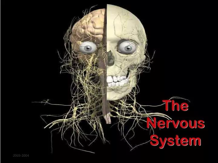
The Nervous System. http://inside.salve.edu/walsh/cns_pns.jpg. Structure and Function of the Neuron. Neuron is the scientific name for a Nerve Cell. Neurons consist of 3 basic structures: Cyton , or cell body. Dendrites- receive messages, impulses, and send them to the cell body.

E N D
Structure and Function of the Neuron • Neuron is the scientific name for a Nerve Cell. • Neurons consist of 3 basic structures: • Cyton, or cell body. • Dendrites- receive messages, impulses, and send them to the cell body. • Axons- send messages away from the cell body. • Nerve impulses travel from one neuron to another across synapses, or spaces in between the cells. • The “jumping across” the synapse is facilitated (helped) by chemicals called Neurotransmitters.
A Neuron Parts of the Cell • Dendrites – Branched parts of a neuron that receive impulses from other neurons. • Cyton- Contains cytoplasm and the nucleus. Impulses pass through here to the axon. • Axon- Single long fiber that carries impulses away from the cell body.
Myelin coating signal direction • Axon coated with insulation made of myelin cells • speeds signal • signal hops from node to node • 330 mph vs. 11 mph myelincoating • Multiple Sclerosis • immune system (T cells) attacks myelin coating • loss of signal
Synapse Junction between nerve cells • 1st cell releases chemical to trigger next cell • where drugs affect nervous system synapse
Depolarization and repolarization. A, RMP results from an excess of positive ions on the outer surface of the plasma membrane. More Na+ ions are on the outside of the membrane than K+ ions are on the inside of the membrane. B, Depolarization of a membrane occurs when Na+ channels open, allowing Na+ to move to an area of lower concentration (and more negative charge) inside the cell-reversing the polarity to an inside-positive state. C, Repolarization of a membrane occurs when K+ channels then open, allowing K+ to move to an area of lower concentration (and more negative charge) outside the cell-reversing the polarity back to an inside-negative state. Each voltmeter records the changing membrane potential as a red line.
Role of ion channels in maintaining the resting membrane potential (RMP). Some K+ channels are open in a "resting" membrane, allowing K+ to diffuse down its concentration gradient (out of the cell) and thus add to the excess of positive ions on the outer surface of the plasma membrane. Diffusion of Na+ in the opposite direction would counteract this effect but is prevented from doing so by closed Na+ channels.
And the answer to how a nerve impulse is conducted… In the nervous system, signals are propagated by action potentials. An action potential is initiated when the dendrites are stimulated, and membrane potential decreases. As the signal moves down the axon of the neuron the voltage-gated sodium channels open, allowing an influx of positive ions. Next, the voltage-gated potassium channels open allowing less positive ions to exit the cell, thus restoring the cell’s negative charge behind the action potential.
Continued… The sodium-potassium pump works all the while to keep the interior of the cell negative by pumping 3 sodium ions out for every 2 potassium ions it pumps in. When the action potential reaches the end of the neuron, the neuron releases neurotransmitters into the synapse. These tiny particles exert their effect on the target cells in one of two ways: They can act to open voltage-gated ion channels by binding directly to the channel, or they can trigger a second messenger when they bind to a cell with a receptor-mediated ion channel.
Types of Neurons Neurons can also be classified by the direction that they send information: ・Sensory (or afferent) neurons: send information from sensory receptors (e.g., in skin, eyes, nose, tongue, ears) TOWARD the central nervous system. ・Motor (or efferent) neurons: send information AWAY from the central nervous system to muscles or glands. ・Interneurons: send information BETWEEN sensory neurons and motor neurons. Most interneurons are located in the central nervous system.
Reflexes • Stimulus- a change in the environment. • Response/Reaction- how the body reacts to a stimulus. • ReflexArc- the pathway that an impulse follows to illicit a response to a stimulus.
What is a reflex? • A reflex is defined as an automatic, involuntary reaction to a stimulus resulting from a nerve impulse passing over a reflex arc. • The reflex arc is an impulse conduction route to and from the central nervous system; it is the smallest part of the nervous system that can receive a stimulus and generate a response.
Types of Reflex Arcs • Ipsilateral • Reflex arc whose receptors and effectors are located on the same side of the body. • Contralateral • Reflex arc whose receptors and efectors are located on opposite sides of the body. • Intersegmental Contralateral • Different sensory receptors deliver stimuli at the same time, and the motor information leaves each segment on the opposite side of the CNS.
With your group… • Think up one scenario that might elicit each type of reflex arc: • Ipsilateral • Contralateral • Intersegmental contralateral
Central Nervous System Peripheral Nervous System Structure of the Nervous System • Major division - Central vs. Peripheral • Central or CNS- brain and spinal cord • Peripheral- nerves connecting CNS to muscles and organs
P e r i p h e r a l N e r v o u s S y s t e m S k e l e t a l A u t o n o m i c ( S o m a t i c ) S y m p a t h e t i c P a r a s y m p a t h e t i c Divisions of the Peripheral Nervous System
Peripheral Nervous System • Connects body to brain & spinal cord • 12 pairs of nerves from your brain (cranial nerves) • 31 pairs from your spinal cord (spinal nerves) • Bundles of sensory and motor neurons held together by connective tissue • Two divisions • Somatic • Autonomic
http://www.christopherreeve.org/Research/Research.cfm?ID=178&c=21http://www.christopherreeve.org/Research/Research.cfm?ID=178&c=21
Divisions of the PNS: Somatic Nervous System • Controls voluntary actions • Made up of the cranial and spinal nerves that go from the central nervous system to your skeletal muscles Autonomic Nervous System • Controls involuntary actions-those not under conscious control-such as your heart rate, breathing, digestion, and glandular functions
Brain Sensory Neuron Motor Neuron Skin receptors Interneuron Muscle Somatic System • Nerves to/from spinal cord • control muscle movements • somatosensory inputs • Both Voluntary and reflex movements • Skeletal Reflexes • simplest is spinal reflex arc
Autonomic System • Two divisions: • sympathetic • Parasympatheitic • Control involuntary functions • heartbeat • blood pressure • respiration • perspiration • digestion • Can be influenced by thought and emotion
CENTRAL NERVOUS SYSTEM SYMPATHETIC Brain Dilates pupil Stimulates salivation Salivary glands Relaxes bronchi Spinal cord Lungs Accelerates heartbeat Heart Inhibits activity Stomach Pancreas Stimulates glucose Liver Adrenal gland Secretion of adrenaline, nonadrenaline Kidney Relaxes bladder Sympathetic ganglia Stimulates ejaculation in male Sympathetic • “ Fight or flight” response • Release adrenaline and noradrenaline • Increases heart rate and blood pressure • Increases blood flow to skeletal muscles • Inhibits digestive functions
Parasympathetic • “ Rest and digest ” system • Calms body to conserve and maintain energy • Lowers heartbeat, breathing rate, blood pressure
Autonomic nervous system controls physiological arousal Sympathetic division (arousing) Parasympathetic division (calming) Pupils dilate EYES Pupils contract Decreases SALVATION Increases Perspires SKIN Dries Increases RESPERATION Decreases Accelerates HEART Slows Inhibits DIGESTION Activates Secrete stress hormones ADRENAL GLANDS Decrease secretion of stress hormones Sympathetic vs. Parasympathetic
Brain Spinal Cord Central Nervous System • Consists of the Brain and Spinal Cord
Central Nervous System • Brain • Spinal cord
Protections • Skull and Vertebrae • 3 protective layers called meninges • Dura Mater (outer layer): consists of connective tissues, blood vessels, and nerves. • Arachnoid Layer (middle layer): elastic and weblike • Pia Mater (inner layer): contains nerves and blood vessels. • Cerebrospinal fluid • a clear watery liquid • separates the middle and inner layers • Acts as shock absorber • exchange of nutrients between blood and nervous system
The Brain • Coordinates body activities • Made up of approximately 100 billion neurons • Uses 20% of bodies oxygen and energy • Divided into three major parts- • the Cerebrum • the Cerebellum • the Brain Stem (Medulla Oblongata, Pons)
Corpus Callosum Right Hemisphere Left Hemisphere Brain has 2 Hemispheres • Left & Right sides are separate • Corpus Callosum : major pathway between hemispheres • Some functions are ‘lateralized’ • language on left • math, music on right • Lateralization is never 100%
Frontal Parietal Occipital Temporal Each hemisphere is divided into 4 lobes
Left visual field Right visual field Optic nerves Left Visual Cortex Corpus Callosum Right Visual Cortex Sensory Information sent to opposite hemisphere • Principle is Contralateral Organization • Sensory data crosses over in pathways leading to the cortex • Visual Crossover • left visual field to right hemisphere • right field to left • Other senses similar
Motor Cortex Somatosensory Cortex Contralateral Motor Control • Movements controled by motor area • Right hemisphere controls left side of body • Left hemisphere controls right side • Motor nerves cross sides in spinal cord
Medial surface of right hemisphere Corpus Callosum Corpus Callosum • Major ( but not only) pathway between sides • Connects comparable structures on each side • Permits data received on one side to be processed in both hemispheres • Aids motor coordination of left and right side
Corpus Callosum • What happens when the corpus callosum is cut? • Sensory inputs are still crossed • Motor outputs are still crossed • Hemispheres can’t exchange data
Verbal left hemisphere Nonverbal right hemisphere The ‘Split Brain’ studies • Surgery for epilepsy : cut the corpus callosum • Roger Sperry, 1960’s • Special apparatus • picture input to just one side of brain • screen blocks objects on table from view
“What did you see?” “Using your left hand, Pick up what you saw.” “What did you see?” ?? I saw an apple. Verbal left hemisphere Nonverbal right hemisphere Verbal left hemisphere Nonverbal right hemisphere The ‘Split Brain’ studies • Picture to left brain • can name the object • left hand cannot identify by touch • Picture to right brain • can’t name the object • left hand can identify by touch
Frontal Parietal Occipital Temporal Localization of Function
Occipital Lobe • Input from Optic nerve • Contains primary visual cortex • most is on surface inside central fissure • Outputs to parietal and temporal lobes Occipital Lobe Visual Lobe
Auditory Cortex Temporal Lobe Temporal Lobe Temporal Lobe • Contains primary auditory cortex • Inputs are auditory, visual patterns • speech recognition • face recognition • word recognition • memory formation • Outputs to limbic System, basal Ganglia, and brainstem
Somatosensory Cortex Parietal Lobe Parietal Lobe • Receives inputs from multiple senses • contains primary somatosensory cortex • borders visual & auditory cortex • Outputs to Frontal lobe • hand-eye coordination • eye movements • attention
Frontal Lobe Working Memory Motor Cortex Motor Cortex Motor Cortex Broca’s Area Frontal Lobe • Contains primary motor cortex • No direct sensory input • Important planning and sequencing areas • Broca’s area for speech • Prefrontal area for working memory
Functions of Different Parts of the Brain • Brainstem • Integration of respiratory and cardiovascular function • Cerebellum • Coordinates Movement • Cerebral Hemispheres • Skilled movement, reason, learning, memory • Thalamus • Control of skeletal-muscle coordination • Awareness • Hypothalamus • Hormones • Balance • Emotions • Thermoregulation • Limbic System • Emotions • Learning
Cerebrum • Largest part of the brain • Thinking • Memory is stored • Movements are controlled • Impulses from the senses are interpreted.
Cerebrum specialization • Regions specialized for different functions • Lobes • frontal • speech, control of emotions • temporal • smell, hearing • occipital • vision • parietal • speech, tastereading parietal frontal occipital temporal