Download

1 / 39
400 likes | 558 Views
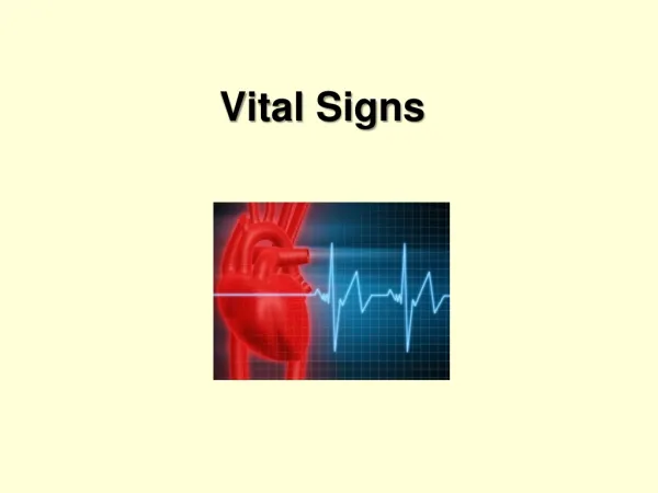
Patient Vital Signs DRAFT. Rad Tech A – Week 13. Patient Interview Role of Radiologic Technologist Elements of the Clinical History. Vital Signs Oxygen Therapy Oxygen Devices Chest Tubes and Lines. Patient Assessment &Vital Signs. Vital Signs. Body Temperature Respiratory Rate

E N D
Patient Vital SignsDRAFT Rad Tech A – Week 13
Patient Interview Role of Radiologic Technologist Elements of the Clinical History Vital Signs Oxygen Therapy Oxygen Devices Chest Tubes and Lines Patient Assessment &Vital Signs
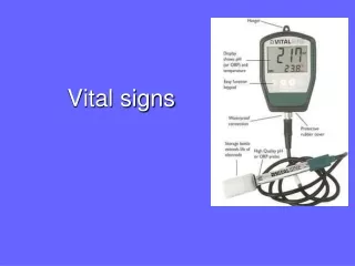
Vital Signs • Body Temperature • Respiratory Rate • Pulse / Heart Rate • Blood Pressure
Indication of Homeostasis Primary Mechanisms Heart beat Blood pressure Body temperature Respiratory rate Electrolyte balance Physical assessment include measurement of vital signs Body Temperature Pulse Respiration Blood Pressure Mental Status Vital Signs
Homeostasis • Our bodies are always trying to maintain HOMEOSTASIS – a constancy in the internal environment of the body, naturally maintained by adaptive responses that promote healthy survival. • Ex: sweating to cool body temperature
Body Temperature • The human body has an ideal temperature, and it works to maintain it, this is called: THERMOREGULATION • Ideal temperature: 98.6 degrees F (oral) • Acceptable range: 97.7 to 99.5 degrees F • Measurement: oral, axillary, tympanic, rectal
Body Temperature • Normal average body temperature: 98.6 F Humans can survive between 106 F and 93.2 F. • Hyperthermia Fever, febrile • Hypothermia below normal range
Measuring Body Temperature • Oral • Rectal • Axillary • Tympanic
Pulse • Pulse rate: Adult = 60 to 100 beats per minute • Children under 10 = 70 to 120 beats per minute • Tachycardia • Bradycardia
Blood Pressure • Measure of the force exerted by blood on the arterial walls during contraction & relaxation. • Measured pressure when the heart is relaxed: Diastolic • Measured pressure when the heart is contracted: Systolic • Measured with a Sphygmomanometer
Blood Pressure • Systolic pressure = 95-140 mmHg • Diastolic pressure = 60-90 mmHg • 120/ 80 Normal
Blood Pressure cont’d • Recorded in millimeters of mercury (mm Hg) with systolic over diastolic • Normal adult systolic: 95-140 mm Hg • Normal adult diastolic: 60-90 mm Hg • Persistent elevation of BP: Hypertension • Persistent low BP: Hypotension
Respiratory Rate • Respiratory System delivers oxygen to the body’s tissues & eliminates carbon dioxide. • Major muscle of ventilation: diaphragm • Measured in “breaths per minute” • Adults: 12 – 20 bpm • Children: 20 – 30 bpm • Newborns: 30 – 60 bpm
Respiratory Rate • Breaths per minute: Adult = 12 to 20 • Children under 10 = 20 to 30 per min • Dyspnea- difficulty breathing • Apnea- no breathing • Bradypnea – decrease is breathing
Pulse Oximeter • Normal Pulse Oximeter = 95% to 100%
Methods of Delivering Oxygen Nasal Cannula Masks Oxyhood Ventilators
Oxygen • Oxygen constitutes 21% of atmospheric gases • If O2 levels in the body drop below 21% homeostasis is altered. • Hypoxia: Inadequate amount of oxygen at the cellular level.
Chest Tubes & LinesThe Rad Tech’s Role • Early detection of problems associated with malpositioned lines. • X-rays assist physicians in determining if tubes and lines are placed correctly • Correct positioning and technical exposure are crucial
Chest Tubes and Lines ENDOTRACHEAL TUBES CHEST TUBES NASOGASTRIC TUBES CENTRAL LINES
Tubes & Lines cont’d • Endotracheal Tubes (ET tubes) – Known as “intubation” -translaryngeal -tracheostomy -nasotracheal • Must be precise in placement: 1-2 inches superior to the tracheal bifurcation (carina)
Intubation of the rt main-stem bronchus with complete occlusion of the lt bronchus causing lt lung atelectasis.
(A) Distal tip of endotracheal tube in rt main bronchus; (B) Central venous catheter in the lt subclavian vein.
Tubes & Lines (last one) • Common insertion sites for CV lines: -subclavian vein -internal jugular vein -femoral vein • Most evaluated by a chest x-ray • Extreme caution must be used when positioning for images!
Rt hydrothorax caused by displacement of a central venous line during dressing change; 1300 ml of intravenous fluids were evacuated via thoracentesis.
Vital Signs Homeostasis Body Temperature Pulse Respiration Blood Pressure Mental Status Electrolyte balance Pulse Oximeter Oxygen Oxygen Devices Chest Tubes Chest Lines Review