Download
1 / 4
40 likes | 77 Views
This study investigates the specificity of two c-Myc Ser62 phosphorylation antibodies from Abcam and Abnova, showcasing their recognition of phosphorylated Ser62 in different cell models. Knockdown of SCP1 is shown to prolong c-Myc half-life, affecting c-Myc stability dependent on FBW7. The research also explores the impact of SCP1 on c-Myc ubiquitination and its role in cell proliferation in a c-Myc dependent manner.

E N D
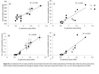
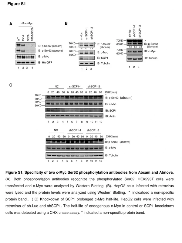
Figure S1 B shSCP1-2 shSCP1-1 NC A HA-c-Myc * (abcam) IB: p-Ser62 shSCP1-1 shSCP1-2 shSCP1-2 shSCP1-1 sh-luc sh-luc IB: c-Myc * * IB: Actin IB: c-Myc T88A/S62A IB: c-Myc * IB: p-Ser62 T58A S62A IB: p-Ser62 IB: p-Ser62 WT IB: Tubulin C IB: c-Myc 1 2 3 4 IB: Tubulin 0 20 40 60 0 20 40 60 0 20 40 60 CHX(min) IB: Tubulin (abnova) IB: p-Ser62 (abcam) IB: p-Ser62 (abnova) IB: c-Myc 60KD 70KD 60KD 70KD 60KD 70KD 60KD 60KD 70KD 60KD 70KD 70KD IB: SCP1 IB: HA-GFP shSCP1-1 NC shSCP1-2 1 2 3 4 5 6 7 8 9 10 11 12 IB: SCP1 0 20 40 60 0 20 40 60 0 20 40 60 CHX(min) (abcam) 1 2 3 1 2 3 (abnova) • Figure S1. Specificity of two c-Myc Ser62 phosphorylation antibodies from Abcam and Abnova. (A). Both phosphorylation antibodies recognize the phosphorylated Ser62. HEK293T cells were transfected and c-Myc were analyzed by Western Blotting. (B). HepG2 cells infected with retrovirus were lysed and the protein levels were analyzed using Western Blotting. * indicatdeda non-specific protein band.. ( C) Knockdown of SCP1 prolonged c-Myc half-life. HepG2 cells were infected with retrovirus of sh-Luc and shSCP1. The half-life of endogenous c-Myc in control or SCP1 knockdown cells was detected using a CHX chase assay. * indicated a non-specific protein band. 1 2 3 4 5 6 7 8 9 10 11 12 1.0 3.1 3.3 1.0 1.8 2.5
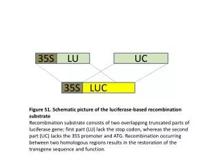
Figure S2 A vector SCP1-WT SCP1-DN Myc(Ub)n IB: c-Myc HA-c-Myc + + + + - - His-Ub - + + + + + FLAG-SCP1 - - WT DN WT DN IB: PP2A B unit IB: SCP1 IB: Tubulin Ni-NTA precipitated 1 2 3 4 5 6 B WCE IB: FLAG D IB: c-Myc C IB: SKP2 IB: SCP1 IB: Actin 1 2 3 4 1 2 3 4 IB: c-Myc siNC siSKP2 IB: c-Myc c-Myc protein expression Figure S2. SCP1 affects c-Myc stability dependent on FBW7. (A). SCP1 has no significant effect on PP2A protein level. HEK293T cells were transfected as indicated. PP2A were analyzed by Western Blotting using PP2A specific antibody (CST #4953) and (B) the different subunits of PP2A were analyzed by QPCR. (C). SCP1 increases c-Myc ubiquitination. HEK293T cells were transfected as indicated. Cells were treated with MG132 for 6 hours, and ubiquitination was analyzed using Ni-NTA-based pull-down assays. (D). Knockdown of FBW7, but not SKP2, blocked the effect of SCP1 on c-Myc stability. HEK293T were transfected with 40nM of control siRNA (siNC), siFBW7 or siSKP2 respectively. After 24h, FLAG-vector or FLAG-SCP1 were transfected into cells in the indicated combinations and analyzed by Western Blotting. siFBW7 siNC SCP1 - + - + SCP1 - + - + IB: FBW7 IB: SCP1 siFBW7 siNC siSKP2 siNC IB: Actin
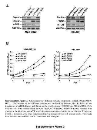
Figure S3 A B IB:c-Myc Bax RhoA D C ** * ** ** ** ** Relative mRNA expression Relative mRNA expression RhoA reporter ** ** ** Relative luciferase activity Figure S3. The transcriptional activity of c-Myc wild-type and its mutant. (A). SCP1 has no significant effect on mRNA level of WDFY2 and PIP5K1A. HEK293T cells were transfected as indicated. The expression of WDFY2 and PIP5K1A mRNA were analyzed by Q-PCR. (B). c-Myc WT, S62A and S62D mediated RhoA reporter gene activity. HEK293T cells were transfected as indicated, the promoter activity were analyzed by Dual-Luciferase assay, c-Myc protein expression were detected by Western blot. (C,D). HEK293T were transfected as indicated, the RNA expression of RhoA or Bax were detected by Q-PCR. The data are means ± s.d. (** P<0.01, t-test; n=3) c-Myc WT c-Myc S62D c-Myc S62A ctrl
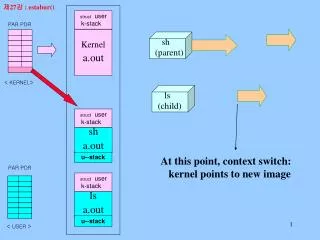
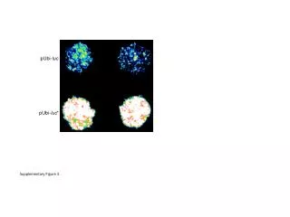
Figure S4 A HeLa 1 2 3 4 OD 490 (nm) B HT29 sh-SCP1 - + - + sh-c-Myc - - + + IB:c-Myc IB: SCP1 IB: Actin 1 2 3 4 OD 490 (nm) Figure S4. SCP1 affects cells proliferation in a c-Myc dependent manner. HeLa (A) or HT29 (B) were infected with retrovirus as indicated. The cell proliferation was detected using MTT assay, and knockdown of c-Myc and SCP1 were analyzed by Western Blotting sh-SCP1 - + - + sh-c-Myc - - + + IB:c-Myc IB: SCP1 IB: Actin